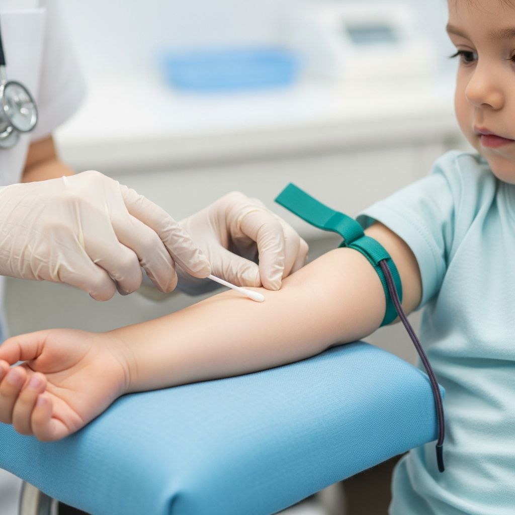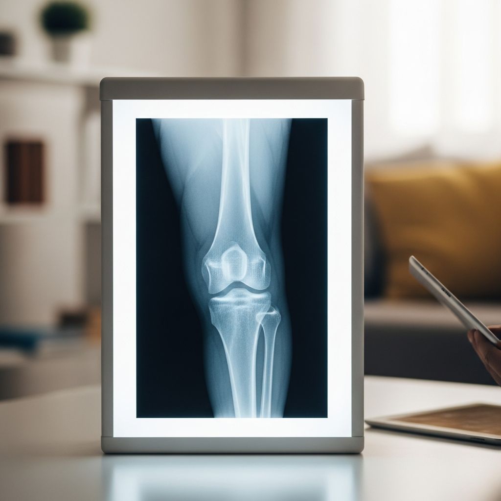
When a child hurts a knee or has long-lasting knee pain, doctors often order a knee X-ray to see what is happening inside the joint. A knee X-ray is a quick, painless imaging test that uses a small amount of radiation to create pictures of the bones in and around the knee.
This guide explains in plain language why a knee X-ray might be needed, what happens before and during the test, how radiation is kept as low as possible for children, and how parents can help their child feel safe and prepared.
Why Doctors Order Knee X-Rays for Children
Doctors do not order X-rays for every sore knee. Instead, they look at your child’s symptoms, how the injury happened, and findings from the physical exam to decide if an image is needed.
Common reasons for a pediatric knee X-ray
- Acute injury from a fall, sports, or accident (for example, a bad twist, collision, or direct blow)
- Inability to walk or bear weight on the leg without significant pain
- Visible deformity of the knee or leg, or the knee looks out of place
- Swelling, bruising, or a large effusion (fluid in the knee) shortly after an injury
- Persistent or unexplained knee pain that does not improve with rest
- Suspected bone disorders, growth plate problems, or developmental conditions
- Checking alignment of the kneecap or bones after a prior injury or surgery
What knee X-rays can help diagnose
A knee X-ray is best at showing bones. It cannot show all types of soft tissue injuries, but it helps rule out many serious bone problems.
- Fractures (broken bones) of the femur, tibia, fibula, or patella
- Growth plate injuries in children and teens, which can affect future bone growth
- Dislocations and subluxations of the knee or kneecap
- Bone tumors or cysts and other unusual bone changes
- Osteochondritis dissecans and other conditions where bone and cartilage may be damaged
- Signs of chronic stress on bone, such as changes at tendon attachment sites
If the X-ray looks normal but the doctor is still concerned about cartilage, ligaments, or other soft tissues, they may order an MRI or ultrasound as a follow-up test.
How a Knee X-Ray Works
A knee X-ray uses a controlled beam of ionizing radiation to create black-and-white images of the knee on a digital detector or film.
- Dense tissues like bone block more X-rays and appear white.
- Softer tissues such as muscle and fat let more X-rays through and look gray.
- Air appears the darkest.
Modern pediatric imaging departments use digital systems that allow radiologists to adjust contrast and brightness, zoom in on problem areas, and measure angles and distances precisely.
Preparing Your Child for a Knee X-Ray
Preparation is simple, but explaining the process in advance can make a big difference in how comfortable your child feels.
Before you arrive
- Explain the test in child-friendly terms, such as “They’re going to take pictures of your knee with a special camera that you can’t feel.”
- Reassure your child that the test does not hurt; the only discomfort usually comes from having to hold still or straighten a sore leg.
- Dress your child comfortably in loose shorts or pants that roll up easily so the knee can be seen.
- Bring comfort items like a favorite toy, stuffed animal, or blanket, especially for younger children.
- Tell the care team about any prior imaging or surgeries on that knee and any metal implants.
Important safety information to share
- Inform the team if your child or teen could be pregnant. Even though knee X-rays use low radiation, the staff may take extra precautions or choose another test.
- Let them know about any difficulty straightening the knee, severe pain, or fear of moving the leg so they can position gently.
- Mention if your child has trouble following directions, high anxiety, or developmental or sensory differences; child-life specialists can help.
What Happens During a Pediatric Knee X-Ray
The actual X-ray usually takes only a few minutes. Expect to be in the radiology department for about 10–20 minutes, including check-in and positioning.
Step-by-step overview
- Check-in and brief history
A staff member will confirm your child’s name and date of birth, ask about the injury, and review any safety questions (such as pregnancy in teens). - Changing or adjusting clothing
Your child may be asked to remove clothing or jewelry from the waist down if these items could block the view. A gown may be provided. - Entering the X-ray room
Inside, there will be an X-ray machine attached to the wall or ceiling and a table or platform for positioning. Parents are usually allowed to stay, wearing a lead apron. - Positioning the knee
A radiologic technologist will help your child lie on a table or stand, depending on the views needed. Pillows, foam blocks, or sandbags may be used to support the leg and keep it still. - Taking the images
The technologist will step behind a protective glass window or into the next room and ask your child to hold still for a few seconds while the picture is taken. Your child will not feel the X-ray beam. - Checking image quality
The technologist will quickly check the images on a screen. If the knee moved or part of the joint is not included, a repeat image may be needed.
Typical knee X-ray views
Several different angles (called views) are usually taken to see the joint clearly and not miss subtle injuries.
| View | How the child is positioned | What it helps show |
|---|---|---|
| Front view (AP) | Leg straight, X-ray beam from front to back | Overall bone alignment, fractures of femur, tibia, fibula, and joint space |
| Side view (lateral) | Lying on side or slightly turned, knee bent | Patella (kneecap) position, joint effusion, subtle fractures |
| Special kneecap view | Knee bent, beam angled toward kneecap | Kneecap alignment and surface details |
Radiation and Safety: What Parents Should Know
Concerns about radiation are normal, especially for children. Health professionals follow strict guidelines to keep exposure as low as reasonably achievable (the ALARA principle), particularly in pediatric imaging.
How much radiation is used?
- A single knee X-ray uses a small dose of radiation, generally much less than imaging tests like CT scans.
- The dose is adjusted for a child’s size and for the specific body part being imaged.
- Newer digital equipment often allows even lower doses than older machines.
Steps taken to protect your child
- Shielding with lead aprons may be used to cover areas such as the abdomen or pelvis when appropriate, especially if parents remain in the room.
- Targeted imaging focuses the beam only on the knee rather than large portions of the leg.
- Minimizing repeat images by careful positioning and clear instructions to hold still.
- Alternatives such as ultrasound or MRI may be recommended if they can answer the question without radiation.
For almost all children, the benefit of an accurate diagnosis from a knee X-ray far outweighs the very small risk associated with the radiation.
After the Knee X-Ray: Results and Next Steps
After the images are taken, your child is free to go home or back to the clinic. There are no activity limits from the X-ray itself; any restrictions come from the knee injury or condition.
How results are interpreted
- A radiologist, a doctor who specializes in reading medical images, reviews the X-rays and writes a formal report.
- The report describes any fractures, alignment issues, growth plate changes, or other abnormalities.
- Your child’s main doctor or emergency provider will pair the X-ray findings with the physical exam and symptoms before explaining the diagnosis.
Possible follow-up plans
- Normal X-ray and improving symptoms: home care, rest, and follow-up only if pain continues.
- Fracture or dislocation: splinting, casting, bracing, or, less commonly, surgery; follow-up X-rays may be needed to monitor healing.
- Growth plate or complex injury: referral to a pediatric orthopedic specialist and possibly advanced imaging (such as MRI or CT).
- Unclear result: repeat X-ray after some time or alternative imaging to clarify the problem.
Helping Your Child Stay Calm and Still
Keeping your child relaxed and still during the X-ray helps get clear pictures and avoids the need for repeat images. Parents can play a major role.
Practical comfort strategies
- Stay close: If allowed, stand near your child’s head or shoulders, wearing a lead apron, and offer your hand to hold.
- Use distraction: Talk about a favorite story, count together, or have your child focus on slow breathing in and out.
- Practice staying still at home by making a game of “statue time” for a few seconds at a time.
- Validate feelings: Acknowledge that it is okay to feel nervous but remind your child that the test is quick and helps the doctor fix what hurts.
- Advocate for breaks if positioning is uncomfortable; the technologist can usually adjust supports or give brief rests between images.
When an X-Ray May Not Be Enough
Some knee problems involve cartilage, ligaments, or small areas of bone damage that can be hard to see clearly on X-ray alone. In those cases, doctors may recommend other imaging tests.
Common additional imaging options
- MRI (Magnetic Resonance Imaging)
Provides detailed pictures of cartilage, ligaments, tendons, and bone marrow without radiation. Often used for suspected ligament tears, meniscus injuries, or osteochondritis dissecans when an X-ray raises concern. - Ultrasound
Uses sound waves (no radiation) to evaluate fluid collections, some soft tissue injuries, or the kneecap in young children. - CT (Computed Tomography)
Combines multiple X-ray slices to give a 3D view of complex fractures or bone shapes. Used more cautiously in children because the radiation dose is higher than with plain X-rays. - Arthrography
Involves injecting contrast dye into the joint followed by X-ray, MRI, or CT to show joint structures more clearly; reserved for specific questions about joint surfaces and stability.
Knee X-Ray FAQs for Parents
Q1: Does a knee X-ray hurt?
No. Your child will not feel the X-ray beam. The only discomfort usually comes from straightening or holding a sore knee still for a few seconds. The technologist will position the leg as gently as possible and can use pillows or supports to make it more comfortable.
Q2: How long does the test take?
The imaging itself normally takes just a few minutes. Including check-in, positioning, and reviewing the pictures, you can expect to be in the radiology area for about 10–20 minutes for a straightforward knee X-ray.
Q3: Is the radiation dangerous for my child?
The radiation dose from a knee X-ray is low and carefully controlled. For most children, the small risk from this dose is far outweighed by the benefit of diagnosing or ruling out a serious injury. Providers follow pediatric radiation safety guidelines and use the lowest dose needed to get a clear image.
Q4: Can I stay in the room with my child?
In many pediatric facilities, a parent or caregiver is encouraged to stay in the room to comfort the child. You will likely be asked to wear a lead apron and stand in a specific area away from the X-ray beam for your own protection.
Q5: When will we get the results?
Timing can vary. In emergency or urgent care settings, a doctor often reviews the X-rays right away and discusses the findings before you leave. In outpatient clinics, a radiologist reads the images and sends a report to your child’s doctor, who then shares the results and next steps with you.
Q6: What if my child cannot hold still?
Technologists experienced with children use gentle supports, quick exposures, and child-friendly explanations to help kids stay still. You can assist by offering comfort and distraction. Sedation is rarely needed for knee X-rays because the test is so quick.
References
- Pediatric Knee Trauma Radiographic Evaluation — Orthobullets. 2023-01-01. https://www.orthobullets.com/pediatrics/322103/pediatric-knee-trauma-radiographic-evaluation
- Knee Pain and Knee Injuries — Children’s Healthcare of Atlanta. 2022-06-01. https://www.choa.org/medical-services/orthopedics/knee-injuries
- Knee X-Ray: Anatomy, Procedure & What to Expect — Cleveland Clinic. 2022-03-29. https://my.clevelandclinic.org/health/diagnostics/23501-knee-x-ray
- Pediatric Arthrography — Children’s National Hospital. 2021-09-15. https://www.childrensnational.org/get-care/health-library/arthrography
- X-Ray Exam — Nicklaus Children’s Hospital. 2020-05-10. https://www.nicklauschildrens.org/treatments/x-ray-exam