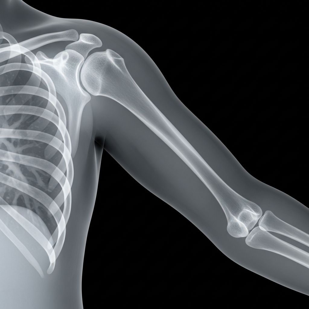
The humerus is the long bone in the upper arm that connects the shoulder to the elbow. When kids fall, collide during sports, or complain of arm pain, healthcare providers often use a humerus X-ray to see what is happening inside the bone and surrounding joints. This guide explains, in simple language, why the test is done, how it works, and how you can help your child feel comfortable throughout the process.
Why Doctors Order a Humerus X-Ray
An X-ray of the humerus gives a detailed picture of the upper arm bone, the shoulder joint, and the elbow area. Providers recommend this test when they need to quickly and safely look for problems that are not visible from the outside.
Common reasons for a humerus X-ray
- Injury after a fall or accident – such as falling off a bike, monkey bars, or during sports.
- Suspected fracture – sudden pain, swelling, or a visible deformity in the arm.
- Shoulder or elbow dislocation – when the joint may have slipped out of place.
- Persistent pain without clear cause – pain that lasts days or weeks, especially with limited movement.
- Monitoring healing – to check whether a broken bone is lining up and healing correctly.
- Checking growth plates – in growing children, the ends of the bone have areas of softer cartilage called growth plates that can be injured.
In emergency situations, a humerus X-ray is usually one of the first tests ordered because it is quick, widely available, and very helpful for making treatment decisions.
How an X-Ray Works (In Kid-Friendly Terms)
An X-ray machine uses a small dose of invisible energy, called X-ray radiation, to make a picture of the inside of the body. Dense structures like bone absorb more of the X-rays and appear white on the image, while softer tissues appear darker.
You can think of it like shining a flashlight through the arm to make a shadow picture on a special digital screen. The radiologic technologist (the imaging specialist running the machine) positions your child’s arm, quickly takes the images, and then the pictures are reviewed by a radiologist and the treating provider.
What the Humerus X-Ray Can Show
A carefully performed humerus X-ray can reveal many different conditions. Often, several views are taken (from the front, side, or angled) so the doctor can see the bone from more than one direction.
| Finding | What It Means | How It May Affect Treatment |
|---|---|---|
| Fracture (broken bone) | A crack or complete break in the humerus. | May require a sling, cast, splint, or sometimes surgery. |
| Dislocation | The humeral head is not sitting correctly in the shoulder joint. | Joint is gently moved back into place, sometimes with sedation, and then supported. |
| Growth plate injury | Damage near the end of the bone where it grows in length. | Needs careful follow-up to be sure growth remains even. |
| Signs of infection or bone disease | Changes in bone shape, density, or structure. | Further tests and specialized treatment may be needed. |
| Arthritis or joint changes | Less common in young children, but can be seen in some conditions. | Guides physical therapy, medication, or other interventions. |
Preparing Your Child for the Test
Good preparation lowers anxiety and helps the exam go quickly and smoothly. Most humerus X-rays are simple outpatient tests that do not require special diet changes or sedation.
Before you arrive
- Explain in simple words: Tell your child that the test will take pictures of their arm bones and that it will not hurt. The hardest part is usually staying still.
- Clothing choices: Dress your child in loose, comfortable clothes that are easy to remove from the upper body. Metal fasteners, zippers, and snaps can show up on the image.
- Remove jewelry: Take off bracelets, watches, necklaces, or other metal objects from the arm, hand, and upper body.
- Talk about previous fears: If your child has been scared during past tests, let the technologist know so they can offer extra reassurance.
Information the care team may ask
- How the injury happened and when symptoms started.
- Whether your child has had any previous fractures or surgeries in the same arm.
- Any known medical conditions or implanted devices.
- In teens who could be pregnant, pregnancy status is important because X-ray use is limited during pregnancy when possible.
Step-by-Step: What Happens During a Humerus X-Ray
Most humerus X-rays take only a few minutes of actual imaging time. Extra time may be needed for check-in and positioning, especially if your child is in pain.
1. Check-in and safety questions
- You and your child will be invited into the imaging department or X-ray room.
- The technologist confirms your child’s identity and the part of the body to be imaged.
- They review safety questions related to pregnancy and metal objects, then help your child remove items that might interfere with the picture.
2. Positioning the arm
- Your child may stand, sit, or lie on the imaging table depending on their age, comfort, and injury.
- The technologist gently positions the arm so both the shoulder and elbow areas are visible.
- Pillows, sandbags, or foam pads might be used to support the arm and keep it as still as possible.
- For a suspected fracture, positioning is done very carefully to avoid worsening pain; the team will ask your child to say if anything hurts.
3. Protection from unnecessary radiation
- Healthcare staff follow the ALARA principle (As Low As Reasonably Achievable), using the smallest radiation dose that will still provide a useful image.
- Lead shields or aprons may be placed over parts of your child’s body not being imaged, when appropriate.
4. Taking the images
- The technologist steps behind a protective barrier or into an adjoining control area.
- Your child will be asked to stay completely still and may be told to hold their breath briefly for each picture to avoid motion blur.
- The machine makes a clicking or buzzing sound, but your child will not feel anything from the X-ray beam.
- Usually, two or more views are taken to allow the doctor to see the humerus from different angles.
5. After the X-ray
- Your child can typically return to normal activities immediately unless their injury requires restriction.
- There is no lingering sensation from the X-ray itself.
- If a serious problem is seen right away, the treating team may discuss the next steps with you while you are still in the facility.
Is the Radiation Safe for Children?
Parents often worry about radiation exposure. Modern X-ray machines are carefully controlled so that children receive only a small, targeted dose. For an upper limb like the humerus, the exposure is relatively low compared with other types of medical imaging.
- Benefits usually outweigh risks: Detecting a fracture or dislocation quickly helps prevent complications and long-term problems.
- Child-sized settings: Pediatric facilities adjust X-ray techniques for a child’s smaller body size to keep doses as low as possible.
- Limited area exposure: The X-ray beam is focused only on the arm, not the whole body.
If you have concerns, ask the radiology team how they minimize radiation and why the study is necessary. They can explain the safety steps used in your child’s case.
How Results Are Interpreted
Once the images are taken, they are typically reviewed by a radiologist—a doctor trained to read medical imaging—and by the clinician treating your child.
What specialists look for
- Bone alignment: Are the pieces of the humerus lined up correctly, or is there a shift, gap, or angle suggesting a displaced fracture?
- Joint position: Does the humeral head sit properly in the shoulder socket and in relation to the elbow joint surfaces?
- Subtle clues: Small changes in bone lines, shadows, or joint spaces can point toward hidden injuries that are not obvious at first glance.
- Soft tissue signs: Swelling around the bone, or certain patterns of fat and fluid in a joint, can signal an unseen fracture.
Sometimes, if the X-ray suggests a complex injury, the doctor may order additional imaging such as a CT scan or MRI for a closer look, especially around the shoulder or elbow joints.
What Happens After the X-Ray?
The next steps depend on what the images show and how your child is feeling. Your child’s age, the exact location of the injury, and whether the bone pieces are in good alignment all help guide treatment decisions.
Possible treatment plans
- No fracture found
Rest, ice, pain relief, and gradual return to activity may be all that is needed. Follow-up is sometimes recommended if pain lasts. - Stable fracture
A sling, splint, or cast may be used. Children’s bones often heal well with proper support because they are still growing. - Displaced or complex fracture
An orthopedic specialist may recommend a more secure immobilization or surgery to realign and stabilize the bone. - Shoulder or elbow dislocation
The joint is usually gently put back into place (reduced), sometimes under sedation or pain medicine, then supported with a sling.
Caring for your child at home
- Follow instructions on how long the sling or cast should stay in place.
- Help your child avoid activities that could lead to another fall.
- Watch for increased pain, numbness, pale fingers, or swelling, and call your provider if these occur.
- Attend all follow-up visits so the team can monitor healing, especially near growth plates.
Tips to Help Your Child Cope
A calm, informed parent is one of the best supports a child can have during any medical test.
- Stay close: When allowed, stand near your child during positioning to offer a hand to hold or reassuring words. You may be asked to wear a lead apron if staying in the room.
- Use distraction: Bring a favorite small toy, story, or song to help them focus on something other than the equipment.
- Practice staying still: Before the visit, play a “statue game” at home to practice holding still for a few seconds at a time.
- Offer choices: Let your child choose which arm rest to use, or what comfort item to bring, so they feel some control.
- Praise their effort: After the exam, highlight how brave and cooperative they were.
Frequently Asked Questions (FAQs)
How long does a humerus X-ray take?
The actual imaging typically takes only a few minutes. Extra time may be needed for check-in, positioning, and any required paperwork, so plan for 20–40 minutes at the facility.
Will my child feel any pain from the X-rays?
The X-ray beam itself is painless. The only discomfort usually comes from holding the arm in one position, especially if there is a fracture or sprain. The team will work gently and adjust positioning as needed.
Can I stay in the room with my child?
In many facilities, a parent or caregiver can stay in the room, provided they wear a protective lead apron and follow staff directions. Some centers may have specific policies, so ask when you arrive.
Are repeat X-rays ever needed?
Yes. Follow-up X-rays are sometimes done to see how a fracture is healing or to check that the bone remains properly aligned as swelling decreases and the child resumes activity.
What if the X-ray is normal but my child still hurts?
Mild injuries, growth plate issues, or soft tissue problems may not always show clearly on an initial X-ray. If pain persists or worsens, your doctor may repeat the X-ray later or order other tests such as ultrasound, CT, or MRI to look more closely.
References
- Shoulder X Ray: Anatomy, Procedure & What to Expect — Cleveland Clinic. 2022-03-01. https://my.clevelandclinic.org/health/diagnostics/22531-shoulder-x-ray
- Proximal Humerus Fractures — Physiopedia. 2023-01-15. https://www.physio-pedia.com/Proximal_Humerus_Fractures
- Shoulder X-ray Interpretation — Don’t Forget the Bubbles. 2020-09-10. https://dontforgetthebubbles.com/shoulder-x-ray-interpretation/
- How to Read an Elbow X-ray — NewYork-Presbyterian. 2019-06-01. https://www.nyp.org/professionals/emergency-medicine/how-to-read-emergency-images/how-to-read-an-elbow-x-ray
- Humerus Series AP and Lateral Views Radiography Positioning — Educational Video, YouTube (academic radiography content). 2018-05-03. https://www.youtube.com/watch?v=TOfgnSxkq0k