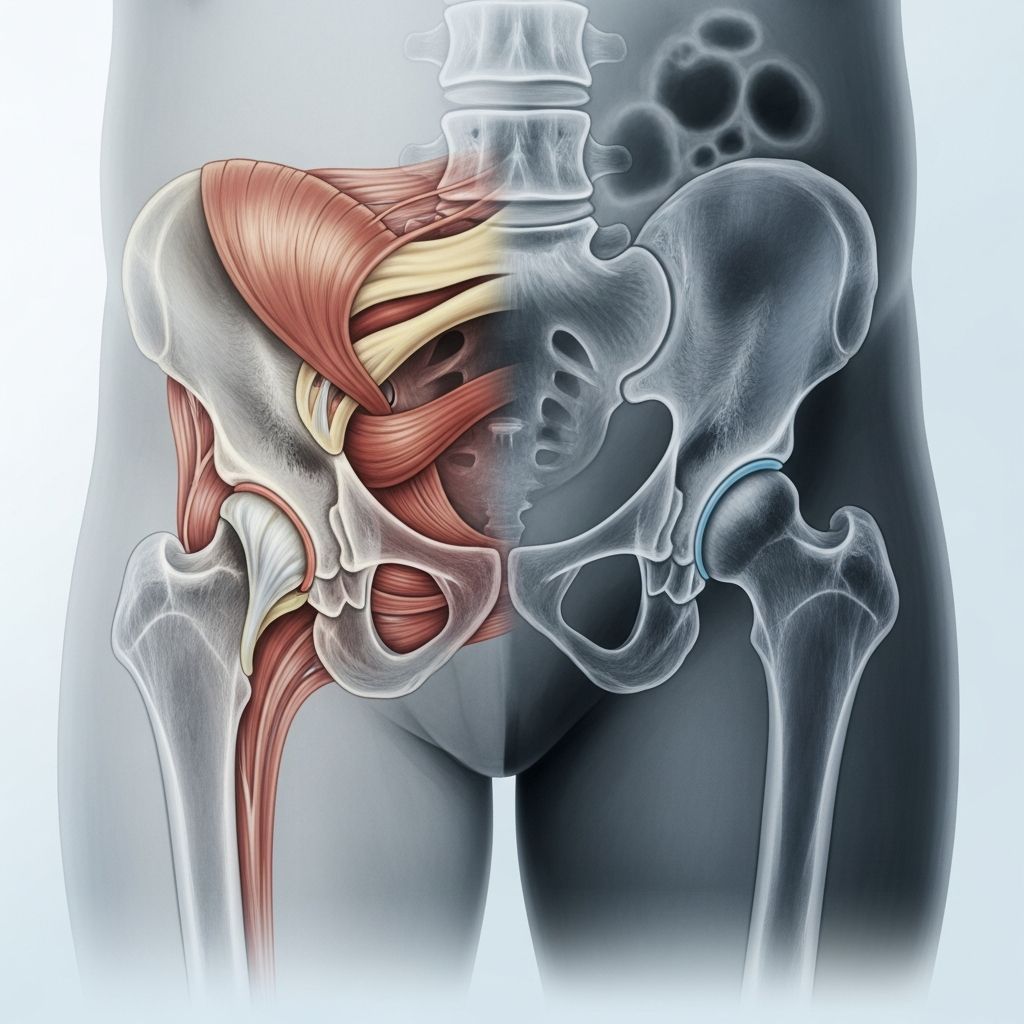
Hip X-rays are a common and safe imaging tool used to evaluate the bones, joints, and surrounding structures in a child’s hip area. These quick procedures help detect issues like developmental dysplasia, fractures, or other abnormalities early, allowing for timely intervention.
Understanding the Basics of Pediatric Hip Imaging
The hip joint connects the thigh bone (femur) to the pelvis, forming a ball-and-socket structure essential for movement and weight-bearing. In children, this joint is still developing, making it susceptible to conditions that X-rays can identify effectively. Unlike ultrasounds used in newborns, X-rays become the preferred method for infants over 4 months and older children because they provide clear views of bone alignment and joint positioning.
X-rays use low-dose radiation to create images of the hip’s bony anatomy. They are particularly useful for assessing the femoral head’s position within the acetabulum (socket) and measuring stability. For growing kids, regular or targeted hip imaging plays a key role in preventing long-term complications like arthritis or dislocation.
Common Reasons for Hip X-Rays in Kids
Pediatricians recommend hip X-rays for various symptoms and risk factors. Pain, limping, or limited range of motion often prompts imaging to rule out fractures, infections, or inflammatory conditions.
- Developmental Dysplasia of the Hip (DDH): This occurs when the hip socket doesn’t fully form, risking dislocation. Breech babies or those with family history are at higher risk; guidelines suggest X-rays at 6 months for breech infants even with normal exams.
- Injuries and Fractures: Falls or trauma can cause pelvic or femoral breaks, visible as disruptions in bone continuity.
- Hip Surveillance in Special Needs: Children with cerebral palsy require routine AP pelvis X-rays to monitor migration percentage, a key metric for dislocation risk.
- Other Conditions: Perthes disease, slipped capital femoral epiphysis, or juvenile idiopathic arthritis may show subtle changes like femoral head collapse or joint space widening.
Early detection through X-rays often avoids surgery; for DDH, braces can reposition the hip while it grows.
How a Hip X-Ray Procedure Works for Children
The process is straightforward and typically lasts just a few minutes. Children lie on an X-ray table, usually face up, with legs in a neutral position—knees straight, feet forward, and pelvis level. Technicians ensure symmetry by checking pelvic wings and obturator foramina on the image.
For optimal results:
- Position the child supine with legs uncrossed and not abducted.
- Use pillows for hip flexion contractures to keep the spine flat.
- A frog-leg lateral view may supplement the standard AP pelvis view for better joint assessment.
Parents can help by explaining the process simply: “You’ll lie still like a superhero while we take a picture of your hips.” Sedation is rare but used for very young or uncooperative kids.
Preparing Your Child for a Hip X-Ray
Preparation is minimal, emphasizing comfort and stillness. Encourage empty bladders to reduce discomfort, and bring a favorite toy or blanket. For toddlers, distraction techniques like stories or videos work well.
| Age Group | Preparation Tips | Expected Duration |
|---|---|---|
| Infants (4-6 months) | Feed beforehand; swaddle if needed | 1-2 minutes |
| Toddlers (1-3 years) | Use play explanations; practice lying still | 2-5 minutes |
| School-age (4+ years) | Explain like a quick photo; reward after | Less than 5 minutes |
No special diet or fasting is required, and metallic objects like jewelry should be removed.
Safety and Radiation Concerns Addressed
X-rays involve minimal radiation, far below levels causing harm, especially with modern digital equipment. The dose for a pelvic X-ray is comparable to background radiation over days, safe even for repeated use in surveillance programs.
Lead shielding for gonads offers negligible benefit and is discouraged due to risks like obscuring anatomy or improper placement. Focus remains on proper positioning to minimize exposure.
Interpreting Hip X-Ray Results
Radiologists analyze key features: femoral head coverage, joint space, bone density, and alignment. In DDH, a dislocated head may reduce with positioning; migration percentage quantifies socket coverage in surveillance.
Normal findings include symmetric hips, round femoral heads, and even joint spaces. Abnormalities like sclerosis or fragmentation suggest Perthes or infection.
- Migration Percentage: Measures uncovered femoral head; over 33% flags intervention.
- H-Line: Horizontal reference for accurate measurements.
Follow-up X-rays track progress during bracing or post-surgery.
Risks, Complications, and When to Worry
Risks are low, but poor positioning can lead to misdiagnosis. Conditions like transient synovitis may mimic serious issues but resolve without imaging intervention.
Seek immediate care for fever, severe pain, or refusal to bear weight, as septic arthritis requires urgent MRI over X-ray.
Treatment Pathways After Hip X-Rays
Results guide care:
- Conservative: Harnesses for DDH in infants.
- Surgical: Arthrograms during operations for precise correction.
- Surveillance: Annual X-rays for at-risk groups like cerebral palsy.
Sweden’s program shows surveillance prevents dislocations population-wide.
Role of X-Rays in Long-Term Hip Health
Consistent imaging from 6 months through adolescence ensures hips develop properly. Breech infants benefit from protocol: ultrasound at 6 weeks, X-ray at 6 months.
For cerebral palsy, proper AP views track growth until triradiate cartilage closes.
Frequently Asked Questions (FAQs)
Is radiation from hip X-rays safe for my baby?
Yes, the dose is very low and safe, equivalent to natural exposure; shielding isn’t needed.
When does my child need a hip X-ray?
For breech babies at 6 months, limping, or DDH risks; consult your pediatrician.
How do I keep my toddler still during the scan?
Use distractions, explanations, or hold hands; it’s quick.
What if the X-ray shows dysplasia?
Bracing often suffices; surgery is rare with early detection.
Are frog-leg views always taken?
Not routinely; added for suspected pathology.
Empowering Parents: Next Steps After Imaging
Discuss results with your orthopedist. Track symptoms and follow recommendations for braces or therapy. Early action preserves mobility for life.
References
- X-ray Screening — International Hip Dysplasia Institute. Accessed 2026. https://hipdysplasia.org/x-ray-screening/
- Hip Dysplasia: X-Ray Recommended for Breech Infants at 6 Months of Age — Seattle Children’s Hospital. 2023-11-01. https://www.seattlechildrens.org/healthcare-professionals/provider-news/hip-dysplasia-x-ray/
- Hip Surveillance Radiograph Tutorial — Shriners Children’s. 2023-10. https://www.shrinerschildrens.org/en/news-and-media/videos/2023/10/hip-surveillance-radiograph-tutorial
- Hip Pathology in Children — The Radiology Assistant. Accessed 2026. https://radiologyassistant.nl/pediatrics/hip/hip-pathology-in-children
- Pelvic X-Ray Exam — Dayton Children’s Hospital. Accessed 2026. https://childrensdayton.org/kidshealth/xray-pelvis/
- Hip X-Ray: Anatomy & Procedure — Cleveland Clinic. Accessed 2026. https://my.clevelandclinic.org/health/diagnostics/23508-hip-x-ray