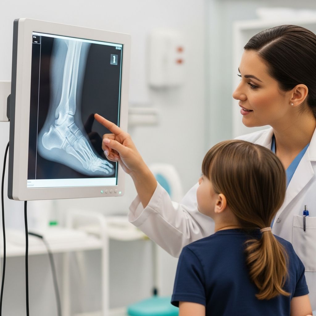
Foot X-rays are a common diagnostic tool used to examine the bones, joints, and soft tissues in a child’s foot, helping doctors identify injuries or conditions causing pain or mobility issues. This imaging test uses low-dose radiation to create clear images, making it quick, painless, and essential for pediatric care.
Understanding the Basics of Pediatric Foot Imaging
The human foot contains 26 bones, including tarsals, metatarsals, and phalanges, along with ligaments and tendons that support movement. In children, these structures are still developing, making them prone to unique injuries from play, sports, or accidents. A foot X-ray produces black-and-white images where dense bones appear white, while softer tissues show as darker shades due to differential radiation absorption.
Unlike adults, kids’ bones are more flexible and heal faster, but growth plates—cartilage areas at bone ends—are vulnerable to damage. X-rays help visualize these without invasive methods, aiding early intervention. Digital X-rays, common today, enhance image clarity and reduce radiation exposure compared to older film methods.
Common Reasons Children Need Foot X-Rays
Parents often seek medical advice when a child limps, complains of foot pain, or shows swelling after a fall. Doctors recommend X-rays for:
- Trauma evaluation: Sprains, fractures, or dislocations from playground mishaps or sports.
- Persistent pain: Unexplained tenderness, especially in heels or toes.
- Swelling or bruising: To check for bone infections, cysts, or fluid buildup.
- Deformities: Assessing alignment issues or growth abnormalities.
- Follow-up care: Monitoring healing after casting or surgery.
Specific conditions include transverse fractures (straight across bones, like Jones fractures), joint dislocations, arthritis signs, heel spurs, or tumors. In pediatrics, X-rays also evaluate post-injury bone positioning even through casts.
Preparing Your Child for a Foot X-Ray
Preparation is minimal, but parental reassurance is key to reduce anxiety. Explain the process simply: “It’s like a quick camera snap of your foot—no needles or pain.” Remove shoes and socks beforehand; metal objects like jewelry should be taken off.
For young children, bring a favorite toy or snack. Facilities often have child-friendly techs who use fun language. No fasting or sedation is needed for standard foot X-rays, unlike more complex scans. If your child has pain, inform the staff—they’ll adjust positioning gently.
| Item | Action |
|---|---|
| Clothing | Wear loose pants; remove shoes/socks |
| Comfort Items | Bring toy, book, or snack |
| Medical History | Share allergies, recent injuries |
| Timing | Arrive 15 mins early |
Step-by-Step: What Happens During the Procedure
A radiologic technologist, trained in pediatric imaging, guides the process. Your child sits or lies on an exam table with the foot positioned on a digital plate. A ceiling-mounted machine captures images.
Typically, three views are taken:
- Anteroposterior (front view): Foot flat, showing bone alignment from above.
- Lateral (side view): Foot turned sideways for depth.
- Oblique (angled view): Rotated 45 degrees to reveal hidden fractures.
Each exposure lasts seconds; total time is under 15 minutes. Stay still is emphasized—techs use verbal cues or gentle holds. A lead apron shields the torso from scatter radiation. No discomfort occurs, though minor repositioning might tickle.
Post-procedure, resume normal activities immediately—no recovery needed. Images are reviewed by a radiologist, often available same-day.
Radiation Safety and Risks for Children
X-rays use ionizing radiation, but pediatric doses are minimized using ALARA (As Low As Reasonably Achievable) principles. A single foot X-ray equals 1-10 days of natural background radiation, far below harmful levels.
Risks are negligible for diagnostic use: no proven cancer link from low-dose exams in kids when justified. Benefits outweigh risks for suspected fractures. Pregnant mothers should inform staff, though foot imaging avoids abdominal exposure. Modern digital tech cuts exposure by 50-90% vs. traditional.
Interpreting the Results: What Doctors Look For
Radiologists analyze images for bone integrity, joint spaces, and soft tissue shadows. Normal feet show uniform bone density; abnormalities include:
- Fracture lines or breaks.
- Displaced bones or widened joints.
- Dark areas indicating cysts or infections.
- Joint narrowing from arthritis or spurs.
Reports detail findings, recommending further tests like MRI if soft tissues need closer look. Orthopedists or podiatrists use results for treatment plans.
When Further Imaging Might Be Needed
X-rays excel at bones but miss some soft tissue issues. Ultrasounds detect ligament tears; CT/MRI for complex fractures or tumors. In kids, follow-ups ensure growth plates heal properly.
Cost, Insurance, and Access for Families
Costs vary: $100-500 uninsured; most pediatric insurances cover fully with referral. Urgent cares or ERs offer quick access; schedule outpatient for non-emergencies.
Tips for Parents Post-X-Ray
- Discuss results promptly with your doctor.
- Follow RICE (Rest, Ice, Compression, Elevation) for injuries.
- Monitor for worsening symptoms.
- Prevent future issues with proper footwear and supervision.
Frequently Asked Questions (FAQs)
Is a foot X-ray painful for my child?
No, it’s painless—just positioning and quick flashes. Kids may feel minor discomfort if injured, but techs accommodate.
How much radiation does it involve?
Very low: equivalent to background exposure. Safe for children with modern equipment.
Can pregnant women stay in the room?
Yes, at a distance; lead shields provided. Inform staff.
What if my child can’t stay still?
Techs use distractions; rare sedation for severe cases.
How soon are results available?
Often same day; radiologist interprets quickly.
Preventing Foot Injuries in Active Kids
Encourage supportive shoes, warm-ups before sports, and safe play areas. Early X-rays catch issues, promoting healthy development.
References
- X-ray of the Foot — Quirónsalud. 2023. https://www.quironsalud.com/en/diagnostic-tests/x-ray-foot
- Foot X Ray: Purpose, Procedure & Risks — Mermaid Beach Radiology. 2024. https://mermaidbeachradiology.com.au/foot-xray-purpose-procedure-risks/
- Foot X Ray Pittsburgh — BV Foot Clinic. 2023. https://bvfootclinic.com/foot-x-ray-pittsburgh/
- Body Parts Foot X-Ray — Scan.com UK. 2024. https://uk.scan.com/body-parts/x-ray/foot
- Diagnostic Studies: X-ray (Radiographs) — 26 Foot & Ankle. 2023. https://26footandankle.com/services/laser-spa-therapies/diagnostic-studies-x-ray-radiographs/
- Foot X-Ray: Anatomy, Procedure & What to Expect — Cleveland Clinic. 2024-01-17. https://my.clevelandclinic.org/health/diagnostics/23500-foot-x-ray