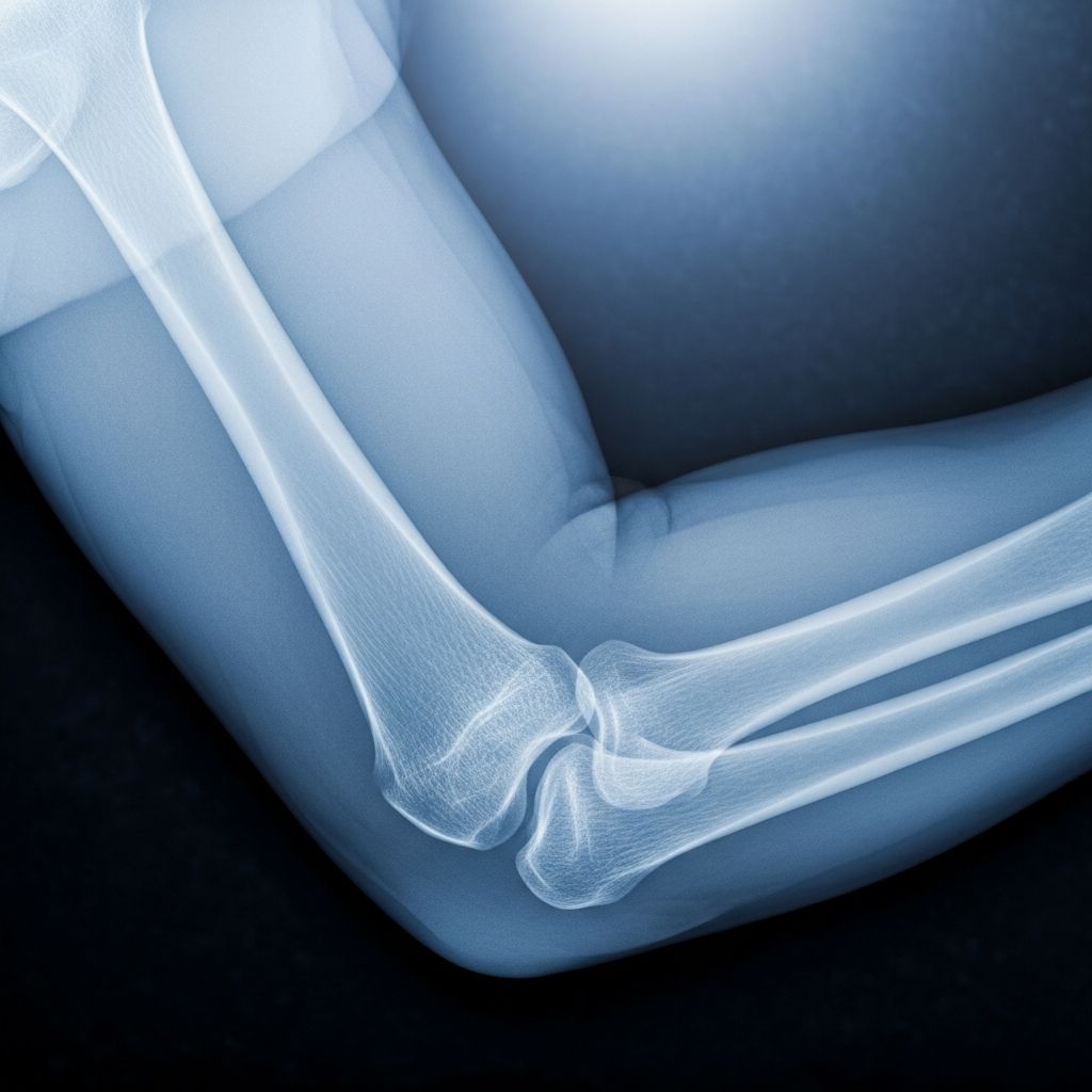
Elbow injuries are common in active children, often from falls or sports. X-rays provide a quick, non-invasive way to visualize bones and joints, helping doctors diagnose fractures or other issues. This guide covers the essentials for parents.
Why Children Need Elbow X-Rays
The elbow joint connects the upper arm bone (humerus) with the forearm bones (radius and ulna). In kids, this area is still developing, with growth plates and cartilage that don’t show up well on standard imaging. Elbow X-rays are crucial after trauma because fractures here make up about 10% of all childhood bone breaks.
Common reasons include falls during play, bike accidents, or sports mishaps. Symptoms like pain, swelling, limited movement, or deformity prompt imaging. Accurate X-rays help detect subtle fractures, especially in young children where bones are mostly cartilage.
Understanding the Developing Elbow Anatomy
A child’s elbow differs from an adult’s due to ossification centers—points where bone forms. These appear in a predictable order remembered by the mnemonic **CRITOE**: Capitellum (1 year), Radial head (5 years), Internal (medial) epicondyle (7 years), Trochlea (9 years), Olecranon (10 years), External (lateral) epicondyle (12 years).
This timeline helps radiologists distinguish normal variants from fractures. For instance, a small bone fragment might be an ossification center, not a break. The cartilaginous structure complicates interpretation, making proper positioning vital.
Standard Positions for Accurate Elbow Imaging
To get clear images, two main views are used: anteroposterior (AP) and lateral. The AP view requires the elbow fully extended with the forearm supinated (palm up). The lateral view needs the elbow flexed at 90 degrees, forearm neutral, and wrist lateral (thumb up).
Challenges arise in injured kids who can’t hold positions due to pain or fear. Studies show only 24% of lateral views are optimal in children, with 63% adequate and 13% suboptimal, often due to rotation or flexion issues.
| View Type | Position Description | Key Challenge in Kids |
|---|---|---|
| Anteroposterior (AP) | Elbow extended, forearm supinated | Full extension painful post-injury |
| Lateral | Elbow 90° flexed, forearm neutral | Rotation from pain or non-compliance |
Innovative Techniques for Better Pediatric X-Rays
Traditional methods use shoulder internal rotation for laterals, but a ‘standing salute’ position—shoulder forward flexed 90°, elbow flexed 90°, forearm neutral—yields truer laterals. In a study of 39 children (average age 4 years), this method better aligned key structures like the ulnohumeral joint and radius-capitellum overlap, reducing repeat exposures.
True lateral signs include: concentric ulnohumeral joint, radial head overlapping capitellum perfectly, and an ‘hourglass’ shape from olecranon and coronoid fossae. Metaphysis and diaphysis widths should match (ratio ~1).
- Salute method benefits: Less rotation, clearer fat pads and lines.
- Reduces radiation: Fewer retakes in uncooperative kids.
Step-by-Step: What Happens During the Procedure
X-rays take 5-15 minutes. A technologist positions the child on the table or against a wall-mounted unit. Parents can stay nearby for comfort, wearing lead aprons.
- Child removes clothing/shirt from injured arm; may wear gown.
- For AP: Lie or stand with elbow straight, palm up.
- For lateral: Bend elbow 90°, thumb up; may use salute pose.
- Hold breath briefly; machine clicks 2-3 times.
- Immobilization aids like sandbags or parental holding if needed.
In emergency rooms, splints may complicate imaging, lowering quality.
Interpreting the X-Ray: Key Signs Doctors Look For
Radiologists use the **FOOL checklist**: Fat pads, Overt findings, Ossification centers, Lines.
- Fat pads: Elevated posterior fat pad signals effusion/fracture (95% specificity).
- Overt findings: Visible breaks, displacements.
- Ossification: Check CRITOE to avoid mistaking centers for fragments.
- Lines: Anterior humeral (bisects capitellum), radiocapitellar (overlaps).
Displacement over 3-4 mm or malalignment suggests epiphyseal fractures needing surgery.
Risks and Radiation Safety Measures
X-rays use low-dose ionizing radiation, safe for kids when minimized. A single elbow series equals 2-3 days of background radiation. Repeat shots increase exposure, so optimal positioning is key.
Alternatives like ultrasound show promise (92% sensitivity for fractures) but aren’t standard everywhere due to operator dependency.
Preparing Your Child for a Comfortable Experience
Explain simply: ‘A quick picture to check your arm.’ Use distractions like toys, videos, or stories. Practice positions at home. Comfort first—technologists accommodate pain.
- Arrive early; bring snacks/comfort items.
- Discuss fears; praise cooperation.
- For toddlers: Parental holding with shielding.
After the X-Ray: Results and Next Steps
Images go to a radiologist immediately. Urgent cases get quick reads. No fracture? Rest, ice, monitor. Fracture? Casting, splinting, or surgery based on type (e.g., supracondylar needs prompt reduction).
Follow-up X-rays track healing. Most heal well with proper care.
Common Myths and Facts About Pediatric Elbow Imaging
| Myth | Fact |
|---|---|
| Always need comparison views of other elbow. | Not routine; adds unnecessary radiation. |
| X-rays hurt. | Painless; discomfort only from positioning. |
| All elbow pain needs X-ray. | Only if swelling, deformity, or limited motion. |
Frequently Asked Questions (FAQs)
Is radiation from elbow X-rays safe for my child?
Yes, doses are minimal and benefits outweigh risks for suspected fractures. Techniques minimize exposure.
How long does an elbow X-ray take?
Usually 5-15 minutes, including positioning.
What if my child can’t stay still?
Technologists use immobilization, distractions, or parental help. Sedation rare for X-rays.
Can ultrasound replace X-rays?
It detects some fractures well but can’t assess bone alignment fully.
When to worry after an X-ray shows no fracture?
Monitor for worsening pain/swelling; follow doctor’s advice.
Empowering Parents: When to Seek Immediate Care
Rush to ER for open wounds, numbness, pale/cold fingers, or severe deformity. Early imaging prevents complications like growth disturbances.
Word count: 1723 (excluding HTML tags and references).
References
- A Practical Method for Obtaining True Lateral Elbow X-rays in Children — NIH/PMC. 2021-02-11. https://pmc.ncbi.nlm.nih.gov/articles/PMC7851228/
- How to Approach the Pediatric Elbow — EMRA. Accessed 2026. https://www.emra.org/emresident/article/pediatric-elbow
- Approach to Pediatric Elbow — Radiology Key. Accessed 2026. https://radiologykey.com/approach-to-pediatric-elbow/
- Variability of Elbow Radiography in the Injured Child — Youth Sports Ortho (PDF). Accessed 2026. https://www.youthsportsortho.com/pdf/variability-in-child-elbow-xrays.pdf
- X-Ray of Pediatric Elbow Fractures — YouTube (C.S. Mott Children’s Hospital). Accessed 2026. https://www.youtube.com/watch?v=qpYhsB89tzU
- Elbow Fractures in Children — Radiology Assistant. Accessed 2026. https://radiologyassistant.nl/pediatrics/hip/fractures-in-children-1
- Pediatric Elbow — University of Utah School of Medicine. Accessed 2026. https://medicine.utah.edu/radiology/education/msk-ref/elbow/pediatric