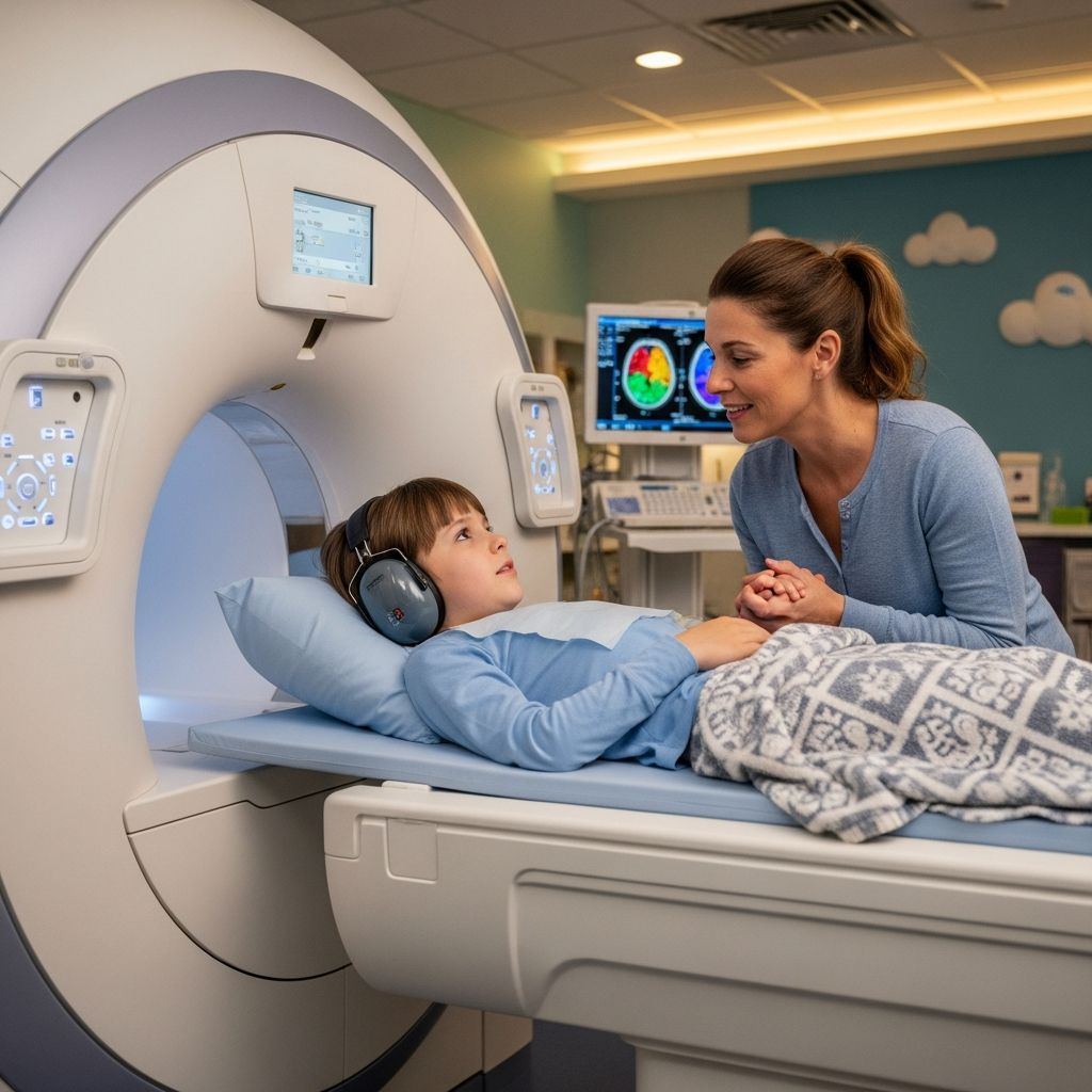
When your child’s doctor recommends a brain magnetic resonance imaging (MRI) scan, you may have questions about what the procedure involves, how to prepare your child, and what happens during the scan. Understanding the process can help reduce anxiety and make the experience smoother for both you and your child. This comprehensive guide provides parents with practical information about pediatric brain MRI scans, preparation strategies, and what to expect at each stage of the procedure.
What Is a Brain MRI and Why Do Children Need It?
A brain MRI is a sophisticated diagnostic imaging technique that uses powerful magnetic fields and radio waves to create detailed three-dimensional pictures of your child’s brain and surrounding tissues. Unlike X-ray or CT scans, MRI does not use ionizing radiation, making it a particularly safe choice for pediatric patients who may require multiple imaging studies over their lifetime. This imaging tool allows physicians to visualize brain structures, identify abnormalities, detect tumors, assess brain development, and monitor conditions affecting the central nervous system.
Doctors may recommend a brain MRI for various reasons, including investigating headaches, evaluating developmental delays, assessing neurological symptoms, following up on previous findings, or monitoring chronic neurological conditions. The detailed images produced by MRI provide information that helps guide diagnosis and treatment planning for your child’s specific medical situation.
How the MRI Machine Works
The MRI scanner is a large, tube-shaped machine that generates a powerful magnetic field around your child’s body. When activated, this magnetic field aligns hydrogen atoms in your child’s tissues. Radio waves are then transmitted through the machine, which causes these atoms to emit signals. A computer detects these signals and converts them into detailed cross-sectional images of the brain from multiple angles. This technology allows radiologists to visualize internal structures with exceptional clarity and precision, revealing details that might not be visible on other types of imaging studies.
The MRI machine’s components work together seamlessly: the large circular magnet creates the magnetic field, radiofrequency coils transmit and receive signals, and gradient coils help locate exactly where signals originate. For brain imaging, a specialized coil may be placed around your child’s head like a helmet to improve image quality and help keep the head still during the scan.
Pre-Appointment Preparation and Planning
Preparation for your child’s brain MRI begins before arriving at the imaging facility. Understanding what to expect and communicating appropriately with your child about the procedure can significantly reduce anxiety and increase the likelihood of a successful scan.
Age-Appropriate Communication Strategies
How you discuss the MRI with your child should match their developmental stage and ability to understand medical concepts. Young children (under 2 years) have limited language comprehension and do best with calm, matter-of-fact approaches focused on sensory experiences. For toddlers and preschoolers (2-4 years), simple, concrete language works best—explain that the machine takes pictures of their brain and that they need to lie still, comparing it to familiar activities they understand. Older children (4-6 years) can grasp more detailed explanations and benefit from understanding why the scan is important for their health, though explanations should remain straightforward and age-appropriate without excessive medical jargon.
Consider scheduling a facility tour if available, as seeing the actual scanner in advance can demystify the experience. Many pediatric imaging centers offer pre-visit videos or virtual tours that help children become familiar with the environment before their appointment.
Practical Pre-Scan Considerations
Review the specific pre-scan instructions provided by your imaging facility, as requirements may vary. Generally, unless instructed otherwise, your child can eat and drink normally before the scan. However, if sedation will be used, your child may need to fast for several hours beforehand—always follow your doctor’s specific instructions on this point.
On the day of the scan, dress your child in comfortable clothing without metal fasteners, zippers, or embellishments. Remove all metal objects before entering the MRI room, including eyeglasses, hearing aids, hairpins, watches, and jewelry. Metal interferes with the magnetic field and can create imaging artifacts or potentially pose safety risks. If your child has any implanted medical devices, inform staff immediately—some devices are not MRI-compatible.
Sedation and Anesthesia Options
One of the most important decisions parents face is whether their child will need sedation or anesthesia during the brain MRI. The need for sedation depends primarily on your child’s age, ability to cooperate, and anxiety level.
When Sedation Is Recommended
Infants and very young children (typically under age 4-5) usually cannot remain still for the 20-90 minute duration required for a brain MRI. Sedation allows young children to sleep through the procedure, eliminating movement that would compromise image quality. Older children who are extremely anxious about enclosed spaces (claustrophobic) may also benefit from sedation to help them remain calm and still during the scan.
Sedation Administration Methods
When sedation is recommended, your child will receive medication either by mouth before arriving at the imaging facility or through an intravenous (IV) line once at the hospital. Oral sedatives are often given 20-30 minutes before the scan to allow time for the medication to take effect. IV sedation provides more controlled dosing and faster onset. A trained medical professional will monitor your child throughout the procedure using equipment that continuously tracks heart rate, breathing, and oxygen levels to ensure safety.
Discussion With Your Medical Team
If your child’s doctor recommends sedation, discuss both the risks and benefits thoroughly. While sedation enables successful imaging by keeping children still, it does carry some risks—though serious complications are rare when administered by experienced professionals in a hospital setting. Your doctor can help you understand whether the diagnostic information gained from the MRI justifies the sedation risks in your child’s specific situation.
What Happens During the Brain MRI Scan
Understanding the step-by-step process of the actual MRI scan helps parents prepare children mentally and know what to expect when arriving at the imaging center.
Arrival and Initial Setup
When you arrive at the imaging facility, staff will review your child’s medical history and any metal implants or devices. You’ll be asked to remove all metal items from your child’s body and clothing. If your child is awake and anxious about the procedure, staff may provide noise-blocking earplugs or headphones before entering the scanner room.
Positioning and Alignment
Your child will lie on a narrow scanning table that slides into the MRI machine. For brain scans, a specialized plastic coil resembling a helmet may be positioned around your child’s head. This coil helps maintain proper alignment, improves image quality, and provides gentle restraint to minimize movement. Once your child is positioned correctly, a red light illuminates to confirm proper alignment with the scanner’s center.
The Scanning Process
The scanning table then slides into the tunnel portion of the MRI machine. If your child is awake, they may or may not be able to see out of the scanner depending on their position and whether a mirror has been positioned to provide an external view. A technologist operates the scanner from an adjacent control room but maintains visual and audio contact with your child through a window and intercom system, allowing two-way communication if needed.
During the actual scanning, the MRI machine produces loud, repetitive banging, knocking, and humming noises as the magnetic field and radiofrequency pulses activate. These sounds are completely normal and indicate the machine is functioning properly. Headphones or earplugs significantly reduce the noise exposure and protect your child’s hearing. Some facilities play music through the headphones to provide distraction and comfort during the scan.
Duration and Movement Requirements
Brain MRI scans typically take 20-90 minutes, depending on the specific imaging protocol needed and the amount of detail required. Throughout the entire scan, your child must lie completely still—even small movements create artifacts that degrade image quality and may necessitate repeating portions of the scan. The technologist will occasionally instruct your child to hold their breath for a few seconds during certain sequences. If your child becomes uncomfortable from lying on the hard table, they may request a blanket for warmth without interrupting the scan.
Contrast Medium Administration
Depending on the clinical question being addressed, your child may receive an injection of contrast medium—a special fluid that helps highlight certain brain structures and abnormalities in the images. The contrast is injected intravenously, usually partway through the scan, and allows radiologists to visualize blood flow, identify inflammation, and detect abnormal tissue enhancement patterns. The contrast medium is safe and typically well-tolerated, though staff will inform you beforehand if it will be used.
After the Scan: What to Expect
Once the scanning is complete, the table slides out of the MRI machine, and your child can sit up. If sedation was used, your child will gradually wake up as the medication wears off. Recovery from sedation typically takes 30 minutes to an hour, during which time trained staff monitor your child until they are fully alert and able to drink and eat normally. You’ll be able to reunite with your child in the recovery area.
Most children can resume normal activities immediately after the scan, though those who received sedation should avoid strenuous activity and operating machinery for the remainder of the day. Follow your imaging facility’s specific discharge instructions, which may include activity restrictions and monitoring guidelines.
Understanding the Results Timeline
After your child’s brain MRI, a radiologist—a physician specialist in medical imaging—reviews all the images carefully and prepares a detailed report. This analysis process takes time, as the radiologist must examine multiple cross-sectional images from various angles to identify any abnormalities or significant findings. The time required varies; complex cases may require consultation with other specialists before results are finalized.
Your imaging facility should provide you with an expected timeframe for receiving results—typically several days to a week. The radiologist’s report is sent to your child’s referring physician, who will discuss the findings with you and explain what they mean for your child’s diagnosis and treatment plan. This physician consultation ensures you understand the results in the context of your child’s specific medical situation.
Reducing Anxiety and Supporting Your Child
Many children experience anxiety about medical procedures, particularly those involving unfamiliar equipment and confined spaces. Several strategies can help minimize fear and support your child through the MRI experience.
- Maintain a calm, matter-of-fact demeanor: Children pick up on parental anxiety, so projecting confidence about the procedure helps your child feel safer
- Use honest, simple language: Explain what will happen in age-appropriate terms without sugar-coating or overwhelming with unnecessary details
- Practice staying still: Play games that reward remaining motionless to help your child develop this skill before the actual scan
- Arrange facility tours: Familiarity with the actual scanner location reduces fear of the unknown
- Bring comfort items: Many facilities allow favorite stuffed animals or blankets in the scanner, providing emotional comfort
- Utilize distraction techniques: Music through headphones, visualizing calming scenes, or counting games can redirect attention from scanner noise
- Discuss the call button: Knowing they can signal for help if distressed provides a sense of control for older children
Frequently Asked Questions About Pediatric Brain MRI
Is an MRI scan safe for children?
Yes, MRI is a very safe diagnostic procedure. It does not use ionizing radiation like X-rays or CT scans, making it particularly suitable for children who may need multiple imaging studies. The magnetic fields and radio waves used pose no known long-term health risks when used appropriately.
Will my child feel pain during the MRI?
No, the MRI scan itself is painless. Your child may feel slight pressure from the coil placed around their head, but this should not cause pain. If an IV line is placed for contrast injection or sedation, there may be minor discomfort from the needle insertion, but this is brief.
Can I stay in the scanner room with my child?
For safety reasons related to the powerful magnetic field, parents typically cannot remain in the scanner room during the actual imaging. However, you can usually stay in a waiting area nearby and can see and communicate with your child through windows and intercom systems. If your child receives sedation, you’ll be reunited in the recovery area.
How long will the appointment take?
The actual brain MRI scan takes 20-90 minutes depending on the specific protocol needed. However, the entire appointment, including check-in, preparation, scanning, and recovery (if sedation is used), typically takes 1.5 to 2.5 hours total.
What should my child wear for the MRI?
Your child should wear comfortable clothing without metal fasteners, zippers, buttons, or metal embellishments. Avoid clothing with metallic threading. Many families find it easiest to change into hospital-provided gowns, which eliminates concerns about metal items.
Why is keeping still so important during the scan?
Even small movements create artifacts—distortions or blurring—in the MRI images that can obscure important details and compromise diagnostic quality. Significant movement may require repeating portions of the scan, extending the procedure time.
What happens if my child can’t stay still during the scan?
If your child cannot remain still without sedation and movement significantly degrades image quality, the technologist may pause the scan and consult with the radiologist and your child’s physician about the best path forward, which might include rescheduling with sedation or using faster imaging protocols if available.
Special Considerations for Different Age Groups
Infants and Young Toddlers (Under 2 Years): This age group is typically scanned while sleeping naturally after feeding and gentle swaddling to minimize movement. Coordination with nap schedules and feeding times optimizes success.
Toddlers and Preschoolers (2-4 Years): As sleep cycles mature, many children in this age range are scanned during their longest continuous sleep period, usually at night. These children need careful preparation and may benefit from sedation if they cannot sleep in the unfamiliar environment.
Older Children (4-6 Years and Beyond): Developing self-regulation capacity allows some older children to cooperate during awake scans, particularly when watching movies or television for distraction. This age group benefits from detailed, honest explanations of the procedure.
Alternative or Complementary Imaging Options
While brain MRI provides exceptional detail for many conditions, other imaging modalities may sometimes be appropriate depending on your child’s specific clinical needs. CT scans provide faster imaging but use radiation. Newer rapid MRI protocols available at some facilities produce diagnostic-quality images in as little as 1-20 minutes—potentially beneficial for anxious children or those unable to tolerate longer procedures.
Your child’s physician can discuss which imaging approach best serves their diagnostic needs, balancing radiation exposure, imaging time, and diagnostic information required.
Preparing for Your Doctor’s Discussion of Results
When your pediatrician or neurologist discusses your child’s MRI results with you, come prepared with questions about what the findings mean, whether additional testing is needed, what treatment options are recommended, and what the expected outcomes are. Request written copies of the report and images if you want to obtain a second opinion or share results with other specialists.
Understanding your child’s brain MRI results allows you to make informed decisions about your child’s healthcare and participate meaningfully in treatment planning with your medical team.
References
- Infant and Child MRI: A Review of Scanning Procedures — National Institutes of Health, National Center for Biotechnology Information. 2021. https://www.ncbi.nlm.nih.gov/pmc/articles/PMC8311184/
- Getting an MRI — Nemours KidsHealth. https://kidshealth.org/en/parents/getting-mri.html
- Making MRIs Less Scary for Kids — Envision Radiology. https://www.envrad.com/making-mris-less-scary-for-kids/
- Pediatric Magnetic Resonance Imaging (MRI) — Children’s National. https://www.childrensnational.org/get-care/health-library/mri
- MRI for Children — Children’s Hospital of Philadelphia. https://www.chop.edu/treatments/magnetic-resonance-imaging-mri
- Scans for Children — The Brain Tumour Charity. https://www.thebraintumourcharity.org/brain-tumour-diagnosis-treatment/how-brain-tumours-are-diagnosed/scans-children/