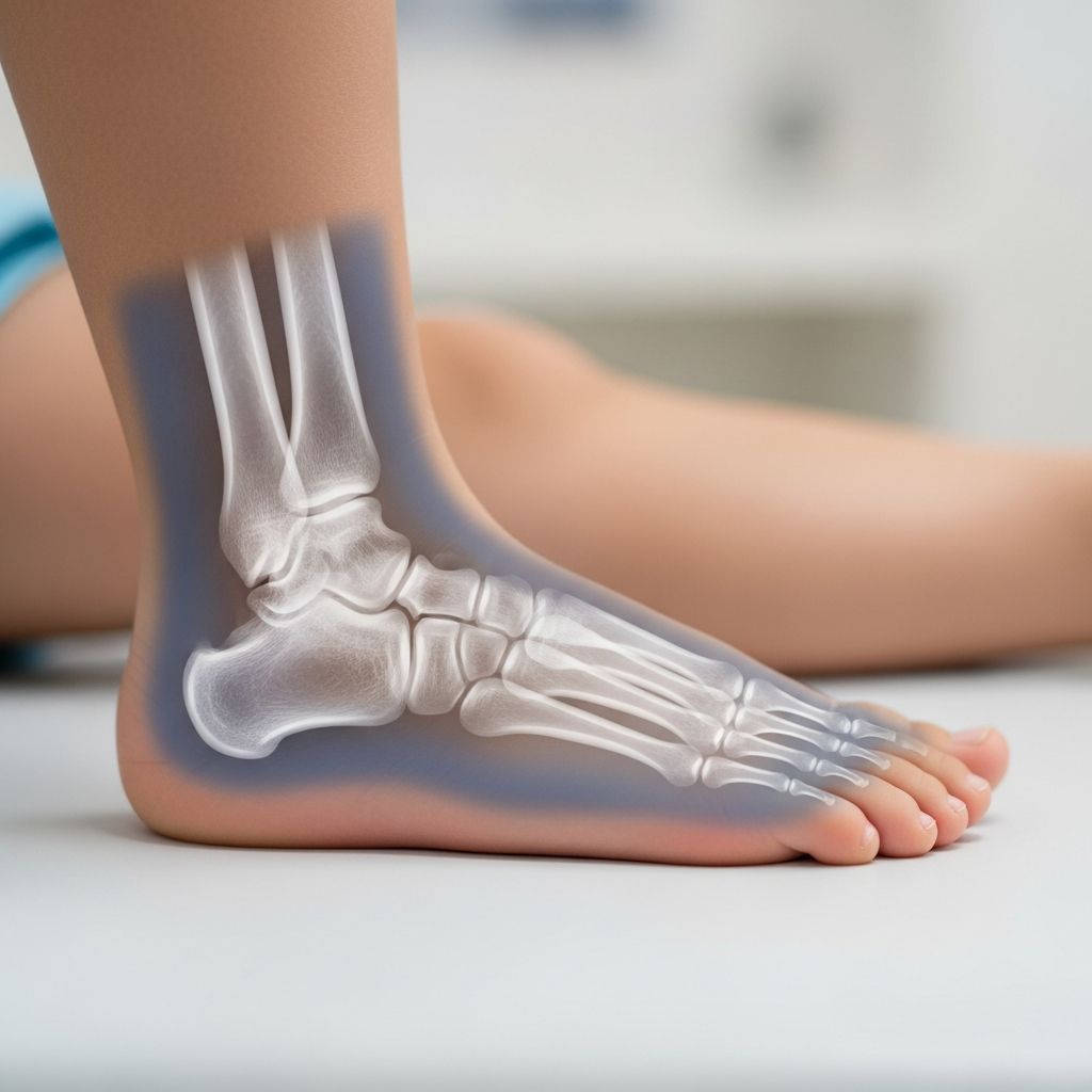
Ankle X-rays are a common diagnostic tool used to evaluate injuries in children’s ankles and feet, helping doctors identify fractures, sprains, or other issues quickly and safely. These low-dose imaging tests provide clear images of bones, guiding treatment decisions from rest to surgery.
Why Children Need Ankle X-Rays
Youngsters are active, often twisting ankles during sports, play, or falls. An ankle X-ray becomes essential when pain, swelling, or bruising suggests bone damage. Unlike adults, kids’ bones are more flexible, so X-rays help distinguish between sprains and fractures that might otherwise be missed.
Common scenarios include playground mishaps, soccer tackles, or bike accidents. Early imaging prevents complications like improper healing, which could affect growth plates—soft areas at bone ends unique to growing children.
Deciding When an X-Ray Is Necessary: Ottawa Ankle Rules
The Ottawa Ankle Rules are evidence-based guidelines to reduce unnecessary X-rays, minimizing radiation exposure for kids. Developed for efficiency, they determine if imaging is needed based on specific exam findings.
For the ankle (malleolar zone):
- Pain in the zone plus bone tenderness at the distal 6 cm of the posterior tibia or medial malleolus tip.
- Pain plus bone tenderness at the distal 6 cm of the posterior fibula or lateral malleolus tip.
- Inability to bear weight immediately and for four steps in the exam room.
For the foot (midfoot zone):
- Pain plus tenderness at the navicular bone base.
- Pain plus tenderness at the fifth metatarsal base.
- Inability to bear weight as above.
These rules boast near 100% sensitivity for fractures, making them ideal for pediatric emergency settings. Parents can ask providers if these criteria apply to avoid overtreatment.
Preparing Your Child for the Procedure
Preparation is simple—no fasting or special diets. Explain the process in age-appropriate terms: “It’s like a quick camera snap of your bones—no pain, just hold still.” Remove shoes, socks, and metal jewelry.
For toddlers, distraction with toys or stories helps. Older kids might watch a video on their device. The whole process takes 10-15 minutes in a radiology suite.
| Age Group | Tips for Cooperation |
|---|---|
| Under 5 | Use parent’s lap, songs, or bubbles. |
| 5-10 | Explain steps, let them practice poses. |
| Over 10 | Involve in positioning, discuss results later. |
How the Ankle X-Ray Is Performed
A radiologic technologist positions the child’s foot on the X-ray plate. Standard views include anteroposterior (AP), mortise (internal rotation 15-20 degrees), and lateral.
- AP View: Foot flat, shows tibia-fibula overlap and joint spaces.
- Mortise View: Foot rotated inward; profiles malleoli, reveals full ankle mortise.
- Lateral View: Side profile; visualizes talus, calcaneus, and fifth metatarsal base.
Weight-bearing views may follow if stable. The machine emits a brief radiation beam, captured digitally for instant review. Technicians ensure distal tibia/fibula and talus are visible for adequacy.
Understanding the X-Ray Images
Images appear black-and-white: bones white, soft tissues gray/black. Doctors check alignment, fractures, and joint spaces.
Key checks:
- Bone Continuity: Trace tibia, fibula, talus for breaks—transverse, oblique, or spiral.
- Joint Spaces: Medial clear space <4mm; tibiofibular overlap >10mm rules out instability.
- Special Angles: Bohler’s angle (20-40°) on lateral for calcaneus integrity.
In kids, look for growth plate injuries (Salter-Harris types), which risk future growth issues.
Common Findings in Pediatric Ankle Injuries
Fractures
Most frequent: lateral malleolus (supination-adduction) or medial (pronation-external rotation). Distal fibula spirals indicate syndesmosis tears.
Sprains and Soft Tissue
No fracture but swelling suggests ligament damage. Joint effusion appears as density changes.
Other Issues
Avulsion fractures (bone chips from ligaments), stress reactions, or foreign bodies. Post-casting X-rays verify alignment.
| Finding | Implication | Treatment |
|---|---|---|
| Clear Fracture Line | Bone break | Cast/boot, possible surgery |
| Normal Bones, Swelling | Sprain | RICE (Rest, Ice, Compression, Elevation) |
| Joint Widening | Instability | Further imaging (CT/MRI) |
Radiation Safety and Risks
X-rays use minimal ionizing radiation, far below harmful levels—equivalent to background exposure over days. Pediatric protocols lower doses via digital tech. Benefits outweigh risks for suspected fractures. Pregnant moms should inform staff.
After the X-Ray: Next Steps
Results are immediate. Normal: home with RICE, crutches if needed. Fracture: casting, boot, or ortho referral. Follow-up X-rays monitor healing, typically 1-2 weeks for kids’ fast bone repair.
Watch for worsening pain, numbness, or color changes—return to ER.
Preventing Ankle Injuries in Active Kids
Strengthen ankles with balance exercises, proper footwear, and warm-ups. Supervise play, teach safe landing from jumps. Sports gear like braces helps high-risk activities.
Frequently Asked Questions
Does an ankle X-ray hurt my child?
No, it’s painless—just positioning. Some discomfort if injured, but quick.
How soon after injury should we get an X-ray?
Immediately if Ottawa rules met; swelling peaks 48 hours, but early imaging is best.
Can kids walk right after?
Usually yes, unless fractured—use crutches if advised.
What if the X-ray is normal but pain persists?
May need MRI for ligaments; follow up with doctor.
Are there alternatives to X-rays?
Ultrasound for soft tissue, but X-ray is gold standard for bones.
Supporting Your Child’s Recovery
Emotional support matters: reassure it’s temporary. Track progress with a journal. Nutrition aids healing—calcium, vitamin D from dairy, veggies. Physical therapy restores strength post-immobilization.
For complex cases like growth plate fractures, pediatric orthopedists specialize in preserving development.
References
- Ottawa Ankle Rules — Physiopedia. 2023. https://www.physio-pedia.com/Ottawa_Ankle_Rules
- Ankle X-ray Interpretation — Geeky Medics. 2023-10-15. https://geekymedics.com/ankle-x-ray-interpretation/
- Foot X-Ray: Anatomy, Procedure & What to Expect — Cleveland Clinic. 2023-05-18. https://my.clevelandclinic.org/health/diagnostics/23500-foot-x-ray
- Ankle x-rays — Don’t Forget the Bubbles. 2022. https://dontforgetthebubbles.com/ankle-x-ray-interpretation/
- Ankle Radiographic Evaluation — StatPearls, NCBI Bookshelf. 2023-07-17. https://www.ncbi.nlm.nih.gov/books/NBK557462/
- Ankle Fracture Mechanism and Radiography — Radiology Assistant. 2023. https://radiologyassistant.nl/musculoskeletal/ankle/fracture-mechanism-and-radiography
- Ottawa Ankle Rules: When to Order an Ankle X-Ray — Medbridge. 2023. https://www.medbridge.com/blog/ottawa-ankle-rules-when-to-order-an-ankle-x-ray