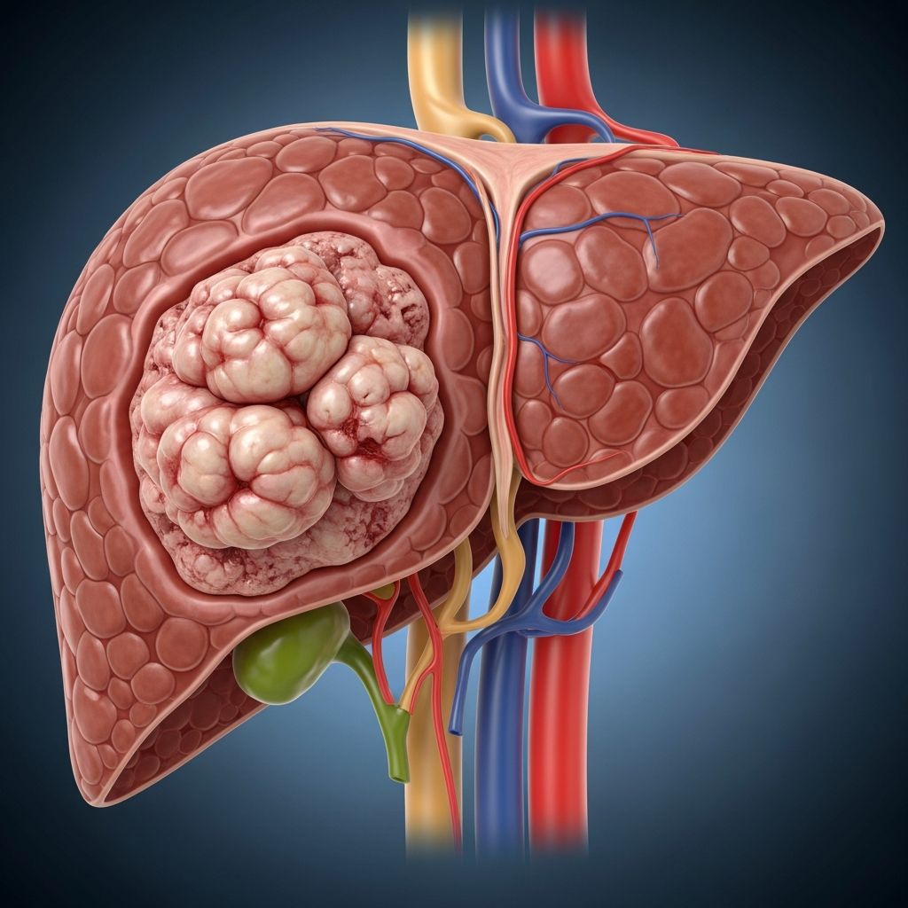
Rare and aggressive, liver rhabdoid tumors primarily affect infants and young children, often presenting as rapidly growing abdominal masses that demand immediate medical attention. These tumors arise from liver cells with genetic alterations, leading to uncontrolled growth and potential spread to other organs.
Recognizing Early Warning Signs
Parents may first notice changes in their child’s behavior or physical appearance due to the tumor’s compressive effects on surrounding tissues. Common indicators include a noticeably distended abdomen, where the belly protrudes outward because of the growing mass. This enlargement can make clothing fit differently or cause visible swelling.
Pain in the abdominal area, particularly on the right side where the liver is located, often accompanies the distension. Children might cry more frequently, refuse to eat, or show discomfort when touched. A palpable mass under the skin can sometimes be felt during routine check-ups or baths, prompting urgent evaluation.
- Yellowing of the skin or eyes, known as jaundice, signals potential liver dysfunction.
- Persistent fever without obvious infection, alongside back pain from tumor pressure on nearby structures.
- Intense itching, possibly due to bile buildup, and sudden loss of appetite leading to unintended weight loss.
- Nausea, vomiting, and anemia manifesting as unusual fatigue or paleness.
These symptoms can escalate quickly, as the tumor grows rapidly in the first year of life. Early detection through vigilant monitoring of these signs can significantly influence outcomes.
Genetic Roots and Risk Factors
At the cellular level, liver rhabdoid tumors stem from disruptions in genes that regulate normal cell division. Healthy cells rely on tumor suppressor genes like SMARCB1 (also called INI1) to prevent uncontrolled proliferation. In these tumors, mutations or deletions in SMARCB1 lead to its loss, allowing malignant cells to multiply unchecked.
Less commonly, alterations in the SMARCA4 gene contribute to tumor formation. These genetic changes are somatic, occurring sporadically rather than being inherited in most cases, though familial patterns are rare. The median age at diagnosis hovers around 15-20 months, with over half of cases appearing before age two.
Incidence remains exceedingly low, estimated at about 1 in 2 million children annually for extra-central nervous system rhabdoid tumors, though underreporting may occur. Infants under one year face higher risks, where these tumors can represent a notable portion of liver malignancies. No strong environmental or lifestyle factors have been definitively linked, emphasizing the role of random genetic events.
Diagnostic Pathways for Confirmation
Diagnosis begins with a thorough physical exam where doctors assess for abdominal masses and related symptoms. Blood tests reveal anemia, elevated liver enzymes like aspartate aminotransferase (AST) and alanine aminotransferase (ALT), and sometimes high alpha-fetoprotein (AFP) levels, though these overlap with other liver tumors like hepatoblastoma.
Imaging plays a pivotal role: Ultrasound detects heterogeneous solid-cystic masses with internal blood flow. MRI shows hypointense T1 and hyperintense T2 signals, fluid-fluid levels indicating hemorrhage, and heterogeneous enhancement. CT scans highlight lobulated, enhancing lesions.
Definitive diagnosis requires biopsy. Pathological examination reveals rhabdoid cells—large cells with eccentric nuclei and eosinophilic inclusions. Immunohistochemistry is crucial: tumors are cytokeratin-positive, INI1-deficient (BAF47 negative), and negative for markers like HepPar-1 or beta-catenin, distinguishing them from hepatoblastoma or other mimics.
| Diagnostic Tool | Key Findings | Purpose |
|---|---|---|
| Ultrasound | Heterogeneous mass, vascularity | Initial screening |
| MRI/CT | T2 hyperintense, hemorrhage | Extent and characteristics |
| Biopsy + IHC | INI1 loss, rhabdoid morphology | Confirmation |
Multimodal Treatment Approaches
Treatment for liver rhabdoid tumors is intensive due to their aggressiveness and propensity for metastasis. A multidisciplinary team—including pediatric oncologists, surgeons, and interventional radiologists—coordinates care.
Neoadjuvant chemotherapy shrinks the tumor pre-surgery. Regimens often include vincristine, doxorubicin, and cyclophosphamide (VAC), sometimes intensified with ifosfamide, carboplatin, and etoposide (ICE). This reduces tumor burden and controls micrometastases.
- Surgical resection aims for complete removal if feasible post-chemo; orthotopic liver transplantation is considered for unresectable cases.
- Embolization manages hemorrhage risks from tumor rupture.
- Radiation therapy targets residual disease, though used cautiously in young children due to long-term effects.
High-dose chemotherapy with autologous stem cell rescue is explored in clinical trials for high-risk cases. Supportive care addresses complications like infections, electrolyte imbalances, and tumor lysis syndrome.
Prognosis and Long-Term Outlook
Despite advances, prognosis remains guarded. Survival rates for localized disease approach 50-60% with aggressive multimodal therapy, but metastatic cases drop below 20%. Factors like age under 6 months, metastasis at diagnosis, and incomplete resection worsen outcomes.
Long-term survivors require vigilant follow-up for secondary cancers, organ dysfunction, and late effects from therapy. Genetic counseling assesses risks for associated rhabdoid predisposition syndromes.
Caring for Your Child Through Diagnosis and Treatment
Navigating a rhabdoid tumor diagnosis is overwhelming. Hospitals often provide child life specialists to ease procedures and psychologists for emotional support. Parents should prioritize nutrition, pain management, and infection prevention.
Join support groups for families facing rare pediatric cancers. Palliative care integrates early to enhance quality of life, addressing symptoms holistically.
Research Frontiers and Hope for Tomorrow
Ongoing trials target SMARCB1-deficient tumors with EZH2 inhibitors and immunotherapy like checkpoint inhibitors. Precision medicine tailors therapies based on molecular profiles, offering promise for better outcomes.
Frequently Asked Questions (FAQs)
What is the most common age for liver rhabdoid tumor diagnosis?
Most cases are diagnosed around 15-20 months, predominantly in infants under two years.
Can liver rhabdoid tumors be cured?
Yes, with complete resection and chemotherapy, some children achieve long-term remission, though relapse risk persists.
How does it differ from hepatoblastoma?
Rhabdoid tumors show INI1 loss on biopsy, poorer prognosis, and rapid growth unlike the more common, often resectable hepatoblastoma.
Is genetic testing recommended for family members?
Yes, if germline SMARCB1 mutations are suspected, to screen siblings for predisposition.
What supportive therapies help during treatment?
Pain relief, anti-nausea meds, nutritional support, and psychosocial counseling are standard.
References
- Rhabdoid Tumor of the Liver (for Parents) — KidsHealth (Humana South Carolina). 2023. https://kidshealth.org/HumanaSouthCarolina/en/parents/rhabdoid-liver.html
- Rhabdoid tumor — Orphanet. 2024-01-15. https://www.orpha.net/en/disease/detail/69077
- Rhabdoid Tumor: Causes, Symptoms & Treatment — Cleveland Clinic. 2024-05-20. https://my.clevelandclinic.org/health/diseases/24440-rhabdoid-tumor
- Pediatric hepatic rhabdoid tumor: A rare cause of abdominal mass — PMC (NCBI). 2018-07-15. https://pmc.ncbi.nlm.nih.gov/articles/PMC6066605/
- Childhood Malignant Rhabdoid Tumor (MRT) — Dana-Farber Cancer Institute. 2024. https://www.dana-farber.org/cancer-care/types/childhood-malignant-rhabdoid-tumor
- Rhabdoid tumor: Symptoms, causes, treatment, and outlook — Medical News Today. 2023-11-10. https://www.medicalnewstoday.com/articles/rhabdoid-tumor-cancer