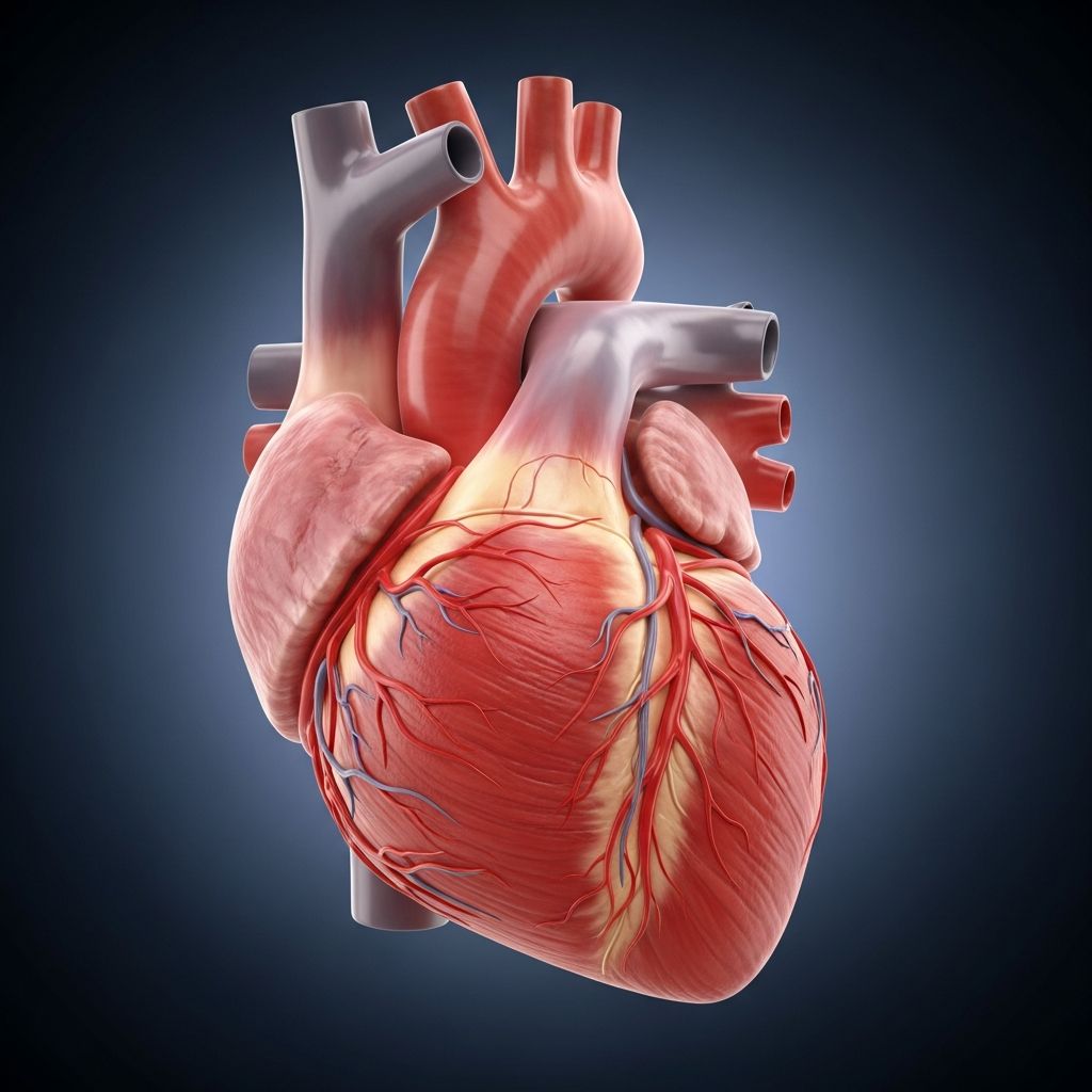
The left ventricle stands as the heart’s most robust chamber, engineered to propel oxygen-rich blood throughout the entire body under immense pressure. This conical muscle drives systemic circulation, distinguishing it from other heart parts by its thickness and strength.
Understanding the Heart’s Layout
The human heart comprises four chambers: two atria on top and two ventricles below. The right ventricle handles deoxygenated blood to the lungs, while the **left ventricle** manages the high-stakes task of systemic distribution. Separated by the interventricular septum—a muscular divider—this design prevents mixing of oxygenated and deoxygenated blood.
Positioned posteriorly and laterally to the right ventricle, the left ventricle forms key heart surfaces: it contributes to the sternocostal area upfront, dominates the diaphragmatic base below, and sharpens into the apex.
Structural Marvels of the Left Ventricle
Shaped like a cone, the left ventricle elongates toward its apex, with walls thickest at the base (up to three times thicker than the right ventricle’s) and tapering to 1-2 mm at the tip. This gradient supports pressure gradients from 120 mmHg systolic peaks.
- External Features: Forms left pulmonary, sternocostal, and diaphragmatic surfaces; base aligns with mitral valve, apex points anteroinferiorly.
- Internal Features: Oval lumen with fine, mesh-like trabeculae near apex; dynamic septum thickens and shortens in systole.
- Valves: Mitral (bicuspid) inlet from left atrium; aortic outlet to systemic arteries.
Papillary muscles—larger anterolateral and posteromedial—anchor chordae tendineae to the mitral valve, resisting systolic pressures to prevent regurgitation.
How Blood Flows Through the Left Ventricle
Oxygenated blood enters from the left atrium via the mitral valve during diastole. Ventricular contraction (systole) seals the mitral and opens the aortic valve, ejecting blood into the aorta.
| Phase | Valve Status | Blood Movement |
|---|---|---|
| Diastole (Filling) | Mitral open, aortic closed | Blood from atrium to ventricle |
| Systole (Ejection) | Mitral closed, aortic open | Blood to aorta and body |
The cardiac cycle relies on electrical impulses from the AV node, bundle of His, bundle branches, and Purkinje fibers to synchronize contraction.
Blood Supply and Venous Return
The left ventricle demands robust perfusion due to its workload. Arterial supply comes primarily from:
- Left anterior descending artery: Free wall and papillary muscles.
- Left circumflex artery: Lateral free wall.
Venous drainage flows via the great cardiac vein, middle cardiac vein, posterior vein of the left ventricle into the coronary sinus, plus Thebesian veins directly into the chamber.
Key Functions and Performance Metrics
Primary role: Generate cardiac output (CO = stroke volume × heart rate) to perfuse organs against systemic resistance. Influenced by preload (venous return), afterload (aortic pressure), and contractility (inotropic state).
Its walls, 3-6 times thicker than the right’s, withstand fivefold stronger forces for body-wide delivery. Efficiency ensures vital organ oxygenation, with output adapting via hypertrophy if needed.
Common Disorders Impacting the Left Ventricle
Left Ventricular Hypertrophy
Chronic pressure overload from hypertension or aortic stenosis causes wall thickening. Initially compensatory, it reduces efficiency, risking failure.
Left Ventricular Failure
Impaired pumping leads to congestion, fatigue, and pulmonary edema. Causes include ischemia, cardiomyopathy, or valvulopathy.
- Symptoms: Shortness of breath, edema, reduced exercise tolerance.
- Risk Factors: High blood pressure, coronary disease, valve issues.
Other Conditions
Atherosclerosis stiffens arteries, demanding more force; aortic valvulopathy exacerbates hypertrophy. Congenital defects may affect children, warranting early screening.
Diagnostic Approaches for Left Ventricle Health
Echocardiography measures ejection fraction (normal 50-70%), wall thickness, and chamber size. ECG detects hypertrophy patterns; MRI offers detailed imaging.
| Test | Purpose | Key Metric |
|---|---|---|
| Echocardiogram | Structure & function | Ejection fraction |
| ECG | Electrical activity | Hypertrophy signs |
| Cardiac MRI | Detailed anatomy | Wall motion |
Signs of Trouble in Children and Adults
Parents should watch for fatigue, poor growth, cyanosis, or murmurs in kids. Adults note dyspnea or chest pain. Early intervention via lifestyle (diet, exercise) and meds prevents progression.
Maintaining Left Ventricle Wellness
Preventive steps include blood pressure control, cholesterol management, regular activity, and avoiding smoking. Annual check-ups aid early detection.
Frequently Asked Questions (FAQs)
What makes the left ventricle thicker than the right?
It pumps against higher systemic pressure, requiring 3-6 times thicker walls.
How does blood enter and exit the left ventricle?
Enters via mitral valve from left atrium; exits through aortic valve to body.
Can left ventricle issues affect children?
Yes, congenital anomalies or acquired conditions like cardiomyopathy can occur; pediatric cardiology monitors these.
What lifestyle changes support left ventricle health?
Balanced diet, exercise, hypertension control, and no smoking reduce risks.
How is left ventricular hypertrophy diagnosed?
Via echocardiogram showing thickened walls and ECG changes.
Why the Left Ventricle Matters for Family Health
As the heart’s engine, its health underpins vitality. Educating families empowers proactive care, from newborn screenings to lifelong habits. Consult specialists for personalized advice.
References
- Heart ventricles: Anatomy, function and clinical aspects — Kenhub. 2023-10-15. https://www.kenhub.com/en/library/anatomy/the-ventricles-of-the-heart
- Left Ventricle Heart — Physiopedia. 2024-05-22. https://www.physio-pedia.com/Left_Ventricle_Heart
- Left Ventricle (Heart) – Function, Definition and Anatomy — YouTube (Anatomy Zone). 2022-11-10. https://www.youtube.com/watch?v=6663FtsNTEc
- Left Ventricle Function, Definition & Anatomy — Healthline. 2023-08-05. https://www.healthline.com/health/left-ventricle
- Left Ventricle — Atlas of Human Cardiac Anatomy, University of Minnesota. 2023-01-12. https://www.vhlab.umn.edu/atlas/left-ventricle/index.shtml
- Heart Anatomy — Texas Heart Institute. 2024-02-28. https://www.texasheart.org/heart-health/heart-information-center/topics/heart-anatomy/