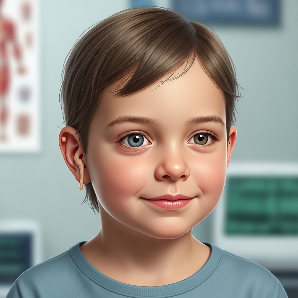
Goldenhar syndrome is a rare congenital condition that primarily impacts the development of facial structures, eyes, ears, and the spine, often presenting asymmetrically on one side of the body. Affecting approximately 1 in 3,000 to 5,000 newborns, it falls under the oculo-auriculo-vertebral spectrum, with varying severity from mild facial differences to complex multi-organ involvement.
Core Characteristics and Physical Manifestations
The hallmark of Goldenhar syndrome involves underdevelopment of bones derived from the first and second branchial arches, leading to distinctive craniofacial features. Children typically exhibit facial asymmetry due to mandibular hypoplasia, where the jaw on one side fails to grow fully, often accompanied by underdeveloped cheekbones.
Eye-related issues are common, including epibulbar dermoids—benign fatty growths on the eye’s surface—microphthalmia (small eyes), or colobomas (notches in the eyelid or iris). Ear anomalies range from preauricular skin tags and microtia (underdeveloped external ears) to anotia (complete absence) and atresia of the ear canal, frequently resulting in hearing impairment.
Spinal deformities such as scoliosis, kyphosis, or fused vertebrae further complicate the picture, potentially affecting posture, mobility, and lung capacity due to rib fusions or absences.
Common Symptoms Breakdown
- Facial Features: Asymmetry, small jaw (micrognathia), wide mouth, cleft lip/palate (in some cases).
- Eye Abnormalities: Dermoids, strabismus, underdeveloped eyes; up to 66% of cases.
- Ear Problems: Microtia/anotia, tags, hearing loss (conductive/sensorineural); seen in 83-100%.
- Skeletal Issues: Vertebral fusions, scoliosis, rib anomalies impacting breathing.
- Other: Heart defects (e.g., septal defects), kidney agenesis, speech/eating difficulties.
While most cases are unilateral, about 10-33% are bilateral, intensifying challenges.
Associated Health Complications
Beyond craniofacial traits, Goldenhar syndrome can involve internal organs. Cardiac anomalies like atrial or ventricular septal defects occur in roughly 50% of cases, necessitating early screening. Renal issues, including unilateral agenesis or multicystic kidneys, affect organ function and require monitoring.
Respiratory concerns arise from airway obstruction due to micrognathia or sleep apnea, while spinal curvatures may limit lung expansion. Neurological impacts are less common but can include developmental delays or intellectual disability in 15% of severe cases, often linked to brain malformations.
| Symptom Category | Approximate Incidence | Source |
|---|---|---|
| Facial Asymmetry/Mandibular Hypoplasia | 65-75% | |
| Ear Anomalies (Microtia, Tags) | 83-100% | |
| Eye Malformations | 30-66% | |
| Vertebral Defects | High (e.g., scoliosis) | |
| Cardiac/Renal Issues | ~50% |
Intellectual function remains typically normal, allowing most children to thrive with interventions.
Diagnostic Approaches and Early Detection
Diagnosis relies on clinical evaluation of characteristic features, often confirmed prenatally via ultrasound detecting facial or spinal anomalies or postnatally through physical exams. Imaging like CT/MRI assesses bone structure, while genetic testing rules out similar syndromes such as Treacher-Collins, differentiated by GS’s asymmetry and less severe malar hypoplasia.
Hearing tests (audiometry), echocardiograms, renal ultrasounds, and ophthalmologic exams form a multidisciplinary baseline. Early identification, ideally in infancy, optimizes outcomes, though some mild cases evade notice until later.
Management and Therapeutic Interventions
Treatment is tailored, multidisciplinary, involving craniofacial surgeons, ENT specialists, orthopedists, cardiologists, and therapists. Surgical corrections for microtia include ear reconstruction using rib cartilage (typically after age 6-10), jaw distraction osteogenesis for asymmetry, and cleft repairs.
- Craniofacial Surgery: Jaw advancement, cheekbone augmentation to improve symmetry and function.
- Ear/Hearing: Bone-anchored hearing aids (BAHA), cochlear implants if needed.
- Eye Care: Dermoid excision, strabismus surgery.
- Spine/Orthopedics: Bracing or spinal fusion for severe scoliosis.
- Supportive: Speech therapy, feeding aids, monitoring for organ issues.
Regular follow-ups track growth, as needs evolve with age. Advances like 3D-printed prosthetics enhance cosmetic and functional results.
Daily Living and Family Support
Families navigate feeding challenges from palate issues or weak facial muscles, often using specialized bottles or gastrostomy tubes temporarily. Speech delays from hearing loss or anatomy prompt early therapy, fostering communication skills.
Psychosocial support addresses self-esteem amid visible differences; counseling and peer groups aid adjustment. Schools may provide IEPs for accommodations like hearing devices or extra time.
Long-term prognosis is positive with care: most achieve independence, though severe cases require ongoing specialist input.
Genetic Insights and Family Planning
Goldenhar syndrome is usually sporadic, not inherited, linked to vascular disruptions or neural crest cell migration errors during weeks 4-8 of gestation. Rare familial cases suggest multifactorial risks; genetic counseling assesses recurrence (low, ~1-2%).
Prenatal care includes anomaly scans; no specific prevention exists, but folic acid may mitigate risks.
Frequently Asked Questions (FAQs)
What causes Goldenhar syndrome?
It arises from disrupted embryonic development of branchial arches, possibly due to vascular issues; not typically genetic.
Is Goldenhar syndrome curable?
No cure, but surgeries and therapies significantly improve appearance, function, and quality of life.
Does it affect intelligence?
Usually not; intelligence is normal in most, with delays rare in severe cases.
When is surgery performed?
Staged: clefts early, jaw/ears in childhood, spine as needed; timing varies by growth.
Can children with Goldenhar lead normal lives?
Yes, with interventions, many attend school, participate in activities, and thrive independently.
Emerging Research and Future Directions
Ongoing studies explore genetic markers via whole-exome sequencing to refine diagnosis and predict severity. Regenerative techniques, like stem cell-based ear reconstruction, promise less invasive options. Multidisciplinary clinics streamline care, improving outcomes.
Parental advocacy groups offer resources; staying informed empowers families.
References
- Goldenhar syndrome – Wikipedia — Wikipedia contributors. 2023-10-15. https://en.wikipedia.org/wiki/Goldenhar_syndrome
- Goldenhar syndrome: clinical features with orofacial emphasis – PMC — PMC. 2013-12-01. https://pmc.ncbi.nlm.nih.gov/articles/PMC3881767/
- Goldenhar Syndrome – Atrium Health Wake Forest Baptist — Wake Forest Baptist Health. 2024-01-01. https://www.wakehealth.edu/condition/g/goldenhar-syndrome
- Oculo-Auriculo-Vertebral Spectrum – Symptoms, Causes, Treatment — NORD. 2023-05-12. https://rarediseases.org/rare-diseases/oculo-auriculo-vertebral-spectrum/
- Goldenhar Syndrome: What It Is, Causes & Symptoms — Cleveland Clinic. 2023-11-20. https://my.clevelandclinic.org/health/diseases/22808-goldenhar-syndrome
- Goldenhar Syndrome Treatment & Diagnose in Dayton, OH | DCH — Children’s Dayton. 2024-02-14. https://childrensdayton.org/conditions/goldenhar-syndrome/