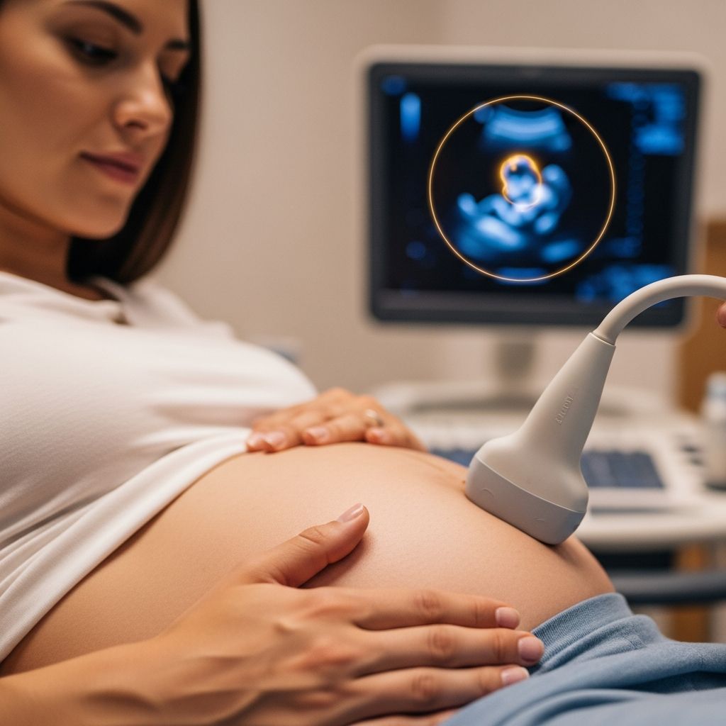
The first ultrasound in pregnancy marks an exciting milestone, offering the earliest glimpse into your developing baby. Typically performed between 6 and 13 weeks, this scan confirms key aspects of early pregnancy development, providing reassurance and critical information for prenatal care.
Why the Initial Pregnancy Scan Matters
Early ultrasound examinations serve multiple vital functions in obstetric care. They establish the pregnancy’s location within the uterus, verify viability through detection of cardiac activity, and determine gestational age for accurate due date calculation. According to clinical protocols, these scans are essential for identifying single or multiple gestations and assessing basic embryonic structures before more advanced development occurs.
Healthcare providers recommend this scan for routine prenatal screening or in response to symptoms like spotting or cramping. It overlaps gynecologic and obstetric evaluations, particularly when confirming intrauterine pregnancy versus ectopic risks.
Optimal Timing for Your Early Scan
Timing is crucial for maximizing the scan’s effectiveness. Scans before 6 weeks may show limited detail, while those after 13 weeks shift focus to second-trimester assessments. Here’s a breakdown of visible milestones by week:
- Weeks 4-5: Gestational sac appears as a small fluid collection, confirming implantation.
- Weeks 5-6: Yolk sac emerges within the sac, supporting early nutrition; fetal pole may be visible.
- Weeks 6-7: Cardiac activity detectable, heartbeat rate 100-170 bpm, rising then stabilizing.
- Weeks 8-10: Embryo measures via crown-rump length (CRL) for precise dating; limb buds form.
- Weeks 11-13: Nuchal translucency screening for aneuploidy; basic anatomy survey begins.
First-trimester dating using CRL is the most accurate, with mean sac diameter (MSD) as backup when no embryo is seen.
Understanding Ultrasound Procedures
Two main approaches exist: transabdominal and transvaginal. Transabdominal uses a gel-covered probe on the abdomen for noninvasive imaging, ideal post-7 weeks. Transvaginal involves a slender probe inserted gently into the vagina for higher resolution in early stages, especially under 7 weeks when abdominal views are obscured by maternal tissue.
Preparation is minimal: arrive with a full bladder for transabdominal scans to enhance uterine visibility. The procedure lasts 15-30 minutes in a darkened room, with real-time images displayed on a monitor.
| Scan Type | Best For | Pros | Cons |
|---|---|---|---|
| Transabdominal | After 7 weeks | Noninvasive, comfortable | Lower resolution early on |
| Transvaginal | Before 7 weeks | Clear early images | Slightly more invasive |
Key Structures Visible in Early Scans
The scan progresses chronologically through developmental landmarks. Initially, the gestational sac—fluid-filled with a mean diameter growing from 2mm at 6 weeks to 6mm by 12 weeks—confirms intrauterine location. The yolk sac, a 3-5mm bubble, follows, connected to the embryo for nutrient transfer.
Next, the fetal pole appears adjacent to the yolk sac, with cardiac flicker visible by 6 weeks. Embryo growth averages 1mm daily, measured as CRL for dating. Heart rate starts at 100-115 bpm pre-6 weeks, peaks at 145-170 bpm by 8 weeks, then plateaus at 137-144 bpm.
Assessing Pregnancy Viability and Health
Viability hinges on cardiac activity and growth progression. Absence of heartbeat with CRL >7mm or no interval growth after 7-10 days indicates non-viable pregnancy. Subchorionic hemorrhage, a common finding, often resolves without issue if small.
Multiple gestations are best diagnosed early; chorionicity (mono- vs. di-chorionic) is determined by membrane thickness and lambda/T-sign, critical for risk assessment.
Dating Your Pregnancy Accurately
Precise dating prevents interventions based on faulty estimates. Primary metrics:
- CRL (6-13 weeks): Gold standard, compared to reference charts.
- MSD: Used pre-embryo visualization.
- BPD (late first trimester): Supplementary measure.
Ultrasound dating trumps last menstrual period (LMP) accuracy, reducing post-term risks.
Spotting Potential Concerns Early
While most scans reassure, anomalies like anembryonic pregnancy (empty sac), blighted ovum, or large yolk sac (>6mm) signal issues. Structural defects such as omphalocele, gastroschisis, or encephalocele may appear post-11 weeks.
Nuchal translucency >3mm prompts further testing for chromosomal issues. Ectopic exclusion is paramount; no sac with rising hCG warrants follow-up.
Benefits of Early Detection
Confirming multiples early allows chorionicity documentation, vital as dichorionic twins have better outcomes than monochorionic. Viability confirmation reduces anxiety, while dating optimizes screening timing like cell-free DNA tests.
Preparing Emotionally and Practically
Expect a mix of excitement and nerves. Bring your partner; discuss results immediately with your provider. Full bladder aids imaging—drink 32oz water 1 hour prior, avoid voiding.
Post-scan, note measurements and heartbeat rate. Follow-up if growth lags or symptoms persist.
Frequently Asked Questions About First Trimester Ultrasounds
Can I see my baby’s heartbeat at the first ultrasound?
Yes, typically by 6-7 weeks via transvaginal scan; rates range 100-170 bpm.
Is it safe to have an early ultrasound?
Absolutely; ultrasound uses sound waves, no radiation, deemed safe by major health organizations.
What if no heartbeat is seen?
Common before 6 weeks; re-scan in 7-10 days. No growth or heartbeat with CRL>7mm suggests non-viability.
How accurate is ultrasound for due dates?
Highly accurate in first trimester (±5 days via CRL), superior to LMP.
Does subchorionic bleeding mean miscarriage?
Not necessarily; many resolve, associated with good outcomes if small and no other issues.
Advances and Future Expectations
Modern scans incorporate 3D/4D for enhanced visualization and AI-assisted measurements for precision. Combined with nuchal translucency and blood tests, first-trimester screening detects 90%+ of Down syndrome cases non-invasively.
Expect personalized care: scans guide lifestyle advice, supplement needs, and monitor high-risk factors like advanced maternal age.
References
- Ultrasound in the First Trimester — Global Library of Women’s Medicine (GLOWM). 2023. https://www.glowm.com/pdf/ultrasound_in_obstetrics_and_gynecology-chapter4.pdf
- Sonography 1st Trimester Assessment, Protocols, and Interpretation — StatPearls, NCBI Bookshelf. 2023-10-01. https://www.ncbi.nlm.nih.gov/books/NBK573070/
- Early Pregnancy Ultrasound: What to Expect at Different Weeks — Roswell OBGYN. 2024. https://roswellobgyn.org/blog/early-pregnancy-ultrasound-what-to-expect-at-different-weeks/
- Patience is key: Understanding the timing of early ultrasounds — UT Southwestern Medical Center. 2023. https://utswmed.org/medblog/patience-key-understanding-timing-early-ultrasounds/
- First Trimester Ultrasound — South Florida Perinatal Medicine. 2024. https://www.sfpm.us/first-trimester-ultrasound.html