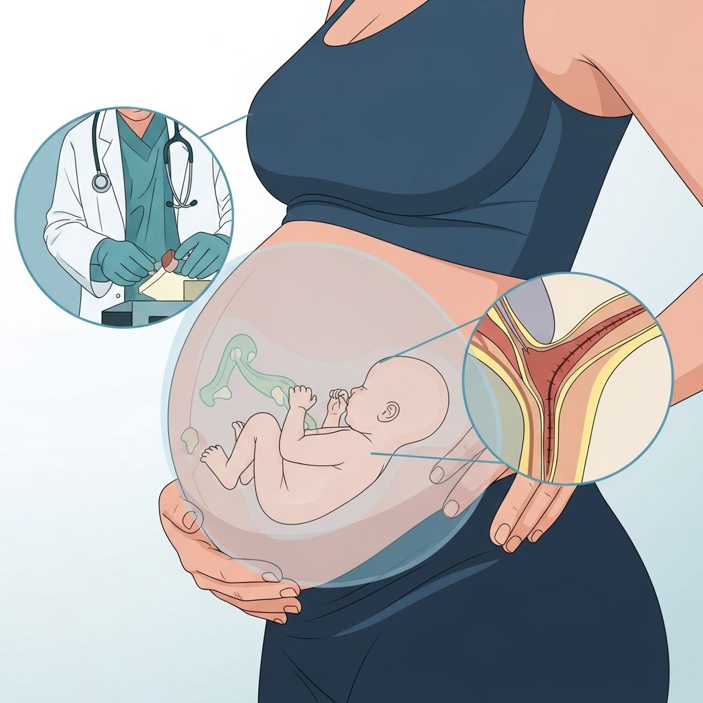
A fetal echocardiogram is a specialized ultrasound that gives doctors a close, detailed look at your unborn baby’s heart. It helps detect heart structure and rhythm problems before birth so that care can be planned early.
What Is a Fetal Echocardiogram?
A fetal echocardiogram (often shortened to fetal echo) uses high-frequency sound waves to create moving images of your baby’s heart while the baby is still in the womb. It focuses on:
- The shape and size of the heart chambers
- Placement and movement of the heart valves
- Blood flow through the heart and major blood vessels
- Your baby’s heart rate and rhythm
Unlike a routine pregnancy ultrasound that checks the whole baby, a fetal echo is dedicated to capturing detailed views of the heart and circulation.
When and Why This Test Is Recommended
Most pregnancies do not require a fetal echocardiogram. Many heart structures can be seen on standard second-trimester ultrasounds. Your healthcare team may suggest this more detailed test when something raises the chance of a heart problem or when a prior scan is unclear.
Typical Timing in Pregnancy
A fetal echocardiogram is most often performed in the second trimester, usually between 18 and 24 weeks of pregnancy. At this stage, the heart is developed enough to be seen clearly, but there is still time to plan care before birth.
In some cases, especially when very early information is needed, a transvaginal (through the vagina) scan may be done in the first trimester to obtain clearer images of the early fetal heart.
Common Reasons Your Doctor Might Order a Fetal Echo
Your obstetrician, midwife, or maternal–fetal medicine specialist may recommend this test if any of the following apply:
- A routine ultrasound suggests an unusual heart structure or an abnormal heartbeat
- There is a family history of congenital heart defects in parents or siblings
- You have certain medical conditions, such as diabetes, lupus, or phenylketonuria
- You took specific medications or were exposed to substances that can affect heart development
- You had infections in early pregnancy that may interfere with heart formation
- The baby shows other structural differences or genetic findings that often occur with heart defects
- Conceived through assisted reproductive technologies, where risk can be slightly increased (depending on the situation)
Sometimes the only reason is that a previous scan did not clearly show the heart. In this situation, the fetal echocardiogram simply provides sharper, more targeted images.
How Fetal Echocardiography Works
The fetal echo uses the same basic technology as routine ultrasound. Sound waves, too high-pitched for you to hear, bounce off your baby’s heart and are converted into images in real time.
| Type | How It Is Done | When It Is Used |
|---|---|---|
| Abdominal fetal echo | Probe (transducer) placed on your belly with gel. | Most common method in the second trimester and later. |
| Transvaginal fetal echo | Smaller probe gently inserted into the vagina. | Sometimes used earlier in pregnancy for clearer images. |
Advanced Views of the Fetal Heart
Depending on your baby’s needs and the center’s technology, the exam may include several techniques:
- 2D echocardiography – provides flat, moving pictures of the heart walls, chambers, and valves
- Doppler echocardiography – shows the speed and direction of blood flow inside the heart and vessels
- Color Doppler – adds color to the Doppler images so that blood flow patterns and any leaks or narrowings are easier to interpret
Step-by-Step: What Happens During the Test
Knowing what to expect can make the appointment feel less stressful. While details vary by hospital or clinic, the basic steps are similar.
Before You Arrive
- You may be asked to wear comfortable, two-piece clothing so your abdomen can be easily accessed.
- Usually, no special diet or fasting is needed, but follow any instructions from your care team.
- Plan to be at the appointment for 45 to 120 minutes, depending on how complex the scan is and how the baby is positioned.
During the Examination
- You lie on a padded exam table, generally on your back or slightly turned to one side.
- A warm, water-based gel is applied to your abdomen to help the sound waves travel.
- The sonographer gently moves the transducer over your belly to capture images from many angles.
- The room is often dimmed so the heart structures are easier for the team to see on the monitor.
- You may be asked to change position or take a deep breath if the baby’s position makes imaging difficult.
The scan is generally not painful, though you may feel mild pressure from the transducer in certain areas.
After the Scan
- The gel is wiped off, and you can return to your normal activities right away.
- A pediatric cardiologist or specially trained physician reviews the images, sometimes while you are still in the clinic.
- You may receive results the same day, or at a follow-up visit, depending on local practice.
Who Performs and Interprets the Test?
A fetal echocardiogram involves a small team of professionals.
- Fetal sonographer or ultrasound technologist – operates the machine and collects images of the heart.
- Pediatric cardiologist – a children’s heart specialist who interprets the images and explains the findings.
- Maternal–fetal medicine specialist or obstetrician – coordinates the test with your overall pregnancy care and discusses next steps.
Is a Fetal Echocardiogram Safe?
Fetal echocardiography uses ultrasound, not radiation. Extensive experience and research show that diagnostic ultrasound, when used appropriately by trained professionals, is considered safe for both mother and baby.
Important safety points:
- The test is noninvasive; there are no needles or injections.
- It is typically painless, apart from mild pressure on the abdomen.
- It does not appear to increase the risk of pregnancy complications.
Professional groups such as the American Institute of Ultrasound in Medicine advise using the lowest ultrasound exposure needed to obtain good images, which is standard practice in fetal echocardiography.
What the Test Can Show
The purpose of the fetal echo is to identify heart conditions as early as possible, so your baby can receive prompt, appropriate care.
Possible Findings
- Normal heart structure and rhythm – the most common result; you continue with routine prenatal care.
- Minor variations – such as small holes between chambers that may close on their own before or after birth.
- Congenital heart defects – structural problems such as narrowed valves, missing or malformed chambers, or incorrectly connected blood vessels.
- Heart rhythm issues – very rapid or slow heartbeats, or irregular rhythms that may need monitoring or treatment.
How Accurate Is the Test?
A fetal echo provides far more detail than a standard obstetric ultrasound and can detect many major congenital heart defects before birth. However:
- Some very small or subtle abnormalities may not be visible prenatally.
- Certain conditions develop or become clearer later in pregnancy or after birth.
- A normal fetal echo greatly lowers, but does not reduce to zero, the chance of a heart problem.
What Happens if a Problem Is Found?
If the test suggests a heart condition, your care team will discuss the findings in detail and create an individualized plan. Early diagnosis allows time to coordinate delivery and treatment at a center equipped for neonatal heart care.
Next Steps May Include
- Additional fetal echocardiograms later in pregnancy to follow how the heart is growing
- Consultation with a pediatric cardiologist to explain the diagnosis and long-term outlook
- Meetings with neonatologists or cardiac surgeons if surgery or special care is expected after birth
- Genetic counseling, especially if heart differences are part of a broader syndrome
- Planning delivery at a hospital with a neonatal intensive care unit (NICU) and pediatric heart team
In some rhythm problems, medications given to the pregnant person can help stabilize the baby’s heartbeat before birth. Management strategies depend on the exact diagnosis and how the baby is doing.
Preparing Emotionally and Practically
Waiting for a fetal echocardiogram and its results can feel stressful. Many parents find it helpful to:
- Bring a partner, family member, or friend for support during the visit
- Write down questions beforehand so they are not forgotten in the moment
- Ask the cardiologist to explain medical terms in everyday language
- Request written information or trustworthy websites for later review
Support groups, counseling services, and hospital social workers can also provide guidance if a heart condition is diagnosed.
Key Takeaways for Parents
- A fetal echocardiogram is a targeted heart ultrasound done during pregnancy to evaluate the baby’s heart in detail.
- It is usually performed between 18 and 24 weeks, and is most often recommended when there is an increased chance of a heart problem or unclear prior scans.
- The test is noninvasive, typically painless, and considered safe for both you and your baby.
- Results help your care team plan the safest possible birth and arrange any heart treatment your baby might need right after delivery.
Frequently Asked Questions (FAQs)
Q1: How is a fetal echocardiogram different from a regular pregnancy ultrasound?
A regular obstetric ultrasound checks the baby’s overall growth and anatomy. A fetal echocardiogram focuses specifically on the heart, using more detailed views and Doppler techniques to evaluate heart structures, blood flow, and rhythm.
Q2: Will my baby feel anything during the test?
No. The test uses sound waves on the outside of your abdomen (or a small probe in the vagina in early pregnancy). There are no injections or instruments touching the baby directly.
Q3: How long does the appointment usually take?
Many fetal echocardiograms take about 45 to 60 minutes, but complex cases or difficult baby positions can extend the exam to as long as two hours.
Q4: If my fetal echocardiogram is normal, does that guarantee a healthy heart?
A normal fetal echo greatly lowers the chance of major congenital heart defects but cannot guarantee that absolutely all heart conditions are absent. Some small or functional issues may only become apparent later in pregnancy or after birth.
Q5: Can I choose not to have the test?
Yes. A fetal echocardiogram is typically recommended, not mandatory. Your healthcare provider should explain the reasons, benefits, and limitations so you can make an informed decision that aligns with your values and preferences.
References
- Fetal echocardiography — MedlinePlus Medical Encyclopedia. 2023-05-15. https://medlineplus.gov/ency/article/007340.htm
- Fetal Echocardiogram Test — American Heart Association. 2022-08-24. https://www.heart.org/en/health-topics/congenital-heart-defects/symptoms–diagnosis-of-congenital-heart-defects/fetal-echocardiogram-test
- Fetal Echocardiography: Purpose, Procedure, and Risks — Healthline. 2022-06-27. https://www.healthline.com/health/fetal-echocardiography
- Fetal Echocardiogram — Children’s Hospital of Philadelphia. 2023-11-02. https://www.chop.edu/treatments/fetal-echocardiogram
- What to Expect During Fetal Echocardiogram — UPMC. 2024-09-05. https://www.upmc.com/services/womens-health/services/tests-procedures/prenatal/fetal-echocardiogram
- Fetal Echocardiography — Lurie Children’s Hospital of Chicago. 2022-10-10. https://www.luriechildrens.org/en/specialties-conditions/fetal-echocardiogram/