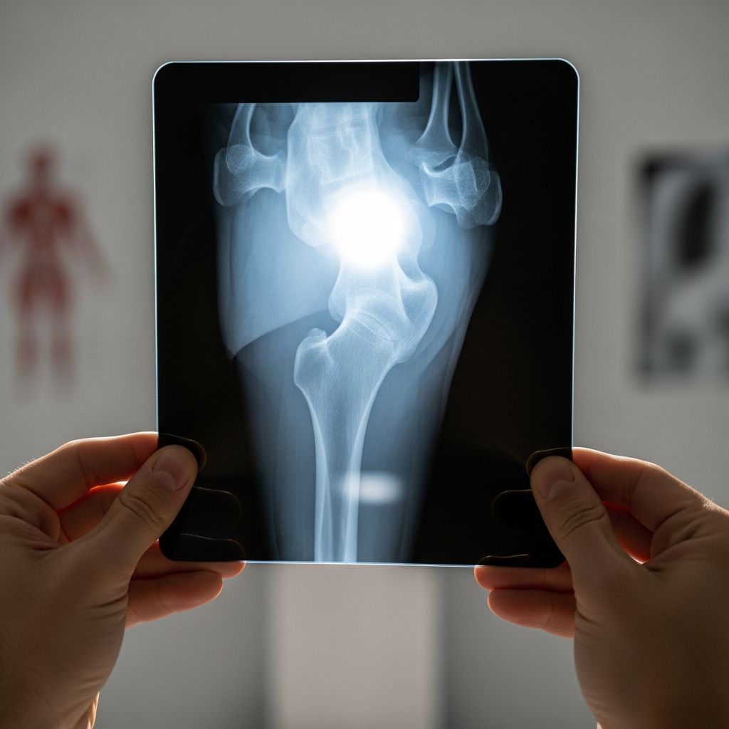
When a child has significant pain or an injury in the upper leg, doctors often recommend an X-ray of the femur, which is the long bone in the thigh. Parents frequently feel anxious about both the injury and the imaging test itself, but understanding the process makes it much easier to handle.
This guide explains, in clear language, why a femur X-ray might be needed, what actually happens in the imaging room, how safe the test is, and how parents can support their child before and after the exam.
What Is the Femur and Why It Matters
The femur is the strongest and one of the longest bones in the body, stretching from the hip joint down to the knee joint. It supports body weight, helps with walking and running, and plays a key role in almost every movement of the lower body.
- The top of the femur forms part of the hip, allowing the leg to move in many directions.
- The bottom of the femur helps form the knee joint, which bends and straightens the leg.
- Muscles, ligaments, and tendons attach along the femur, contributing to strength, balance, and stability.
Because the femur is so important for movement and weight-bearing, any injury or abnormality in this bone can affect how a child walks, runs, or even stands.
What a Femur X-Ray Actually Is
A femur X-ray is a simple imaging test that uses a controlled amount of radiation to create pictures of the thighbone and the surrounding area. The images show the bone’s shape, alignment, and density, and can reveal a variety of problems.
During the exam, an X-ray machine sends an invisible beam through the upper leg toward a detector (either digital or film-based) on the opposite side. Different tissues absorb the beam in different amounts, producing a black-and-white image that highlights bone structures much more clearly than soft tissues.
- Noninvasive: No needles or incisions are involved.
- Quick: The actual exposure takes less than a second, though positioning can take a few minutes.
- Painless: The machine does not touch or hurt the skin, although lying still may be uncomfortable if the leg is injured.
Common Reasons Doctors Order a Femur X-Ray
Doctors order a femur X-ray when symptoms or physical exam findings suggest a problem with the thighbone or nearby joints. The goal is to see what is happening inside the leg and guide treatment decisions.
Typical symptoms that prompt imaging
- Sudden or severe pain in the upper leg or thigh
- Visible deformity, such as the leg looking crooked or shorter
- Inability or refusal to walk, stand, or put weight on the leg
- Swelling, bruising, or warmth around the thigh area
- Unexplained limping or change in walking pattern
Conditions a femur X-ray can help detect
- Broken bones (fractures): Cracks or complete breaks in the femur, sometimes with pieces shifted out of alignment.
- Growth plate injuries: Damage near the ends of the bone where new bone forms as a child grows.
- Bone infections: Areas that suggest infection within the bone.
- Abnormal growths: Unusual bone lumps or areas that might represent benign or more serious conditions.
- Post-injury healing: Follow-up views to see whether a previously broken femur is healing properly.
How to Prepare Your Child for a Femur X-Ray
Most femur X-rays require very little preparation, but planning ahead can help the visit go smoothly and reduce your child’s anxiety. You do not need to change your child’s diet or stop regular medications unless the doctor gives specific instructions for another reason.
What to do before the appointment
- Dress your child in comfortable clothing, ideally without metal snaps, zippers, or thick logos over the leg area.
- Bring any previous imaging reports or discs if they were done at another facility.
- Pack a favorite small toy, stuffed animal, or book to provide comfort in the waiting area.
- Arrive a bit early to fill out paperwork and avoid feeling rushed.
How to talk to your child
- Use simple, reassuring language such as “The doctor will take pictures of your leg so we can see how it’s doing.”
- Emphasize that the machine will not hurt and that the hardest part is staying as still as a statue for a few seconds.
- For young children, turn it into a game: who can stay the stillest during the picture?
- Answer questions honestly but calmly, focusing on safety and comfort.
Step-by-Step: What Happens During the Exam
Knowing what to expect in the imaging room helps parents feel more in control and better able to support their child. While every hospital or clinic has its own flow, most femur X-ray exams follow similar steps.
Arrival and check-in
- You and your child will check in at the imaging or radiology department desk.
- A staff member may ask about your child’s symptoms, medical history, and any prior imaging.
- Depending on the facility, your child may change into a gown, especially if clothing has metal over the area being imaged.
Positioning in the X-ray room
- A radiologic technologist (a trained imaging professional) will greet you and explain the process.
- Your child will be helped onto a table or positioned standing, depending on age, injury, and the views needed.
- The technologist will gently move the leg into the right position and may place foam pads or sandbags to keep it still.
- Parents are often allowed to stay in the room, wearing a lead apron, to comfort the child and encourage stillness.
Taking the images
- For most femur studies, images are taken from at least two angles (for example, from the front and from the side).
- The X-ray tube may move above or beside your child, but it does not touch the body.
- The technologist will step back behind a protective barrier or into an adjacent area while activating the X-ray.
- Your child will be asked to hold very still and may be asked to hold their breath for just a second or two to avoid blurring.
The actual exposure lasts only a fraction of a second, but with positioning and multiple views, the entire exam typically takes several minutes. If the child moves and an image is blurry, an extra picture may be necessary.
Radiation Exposure and Safety
One of the most common parental concerns is radiation. It is important to know that modern X-ray equipment uses the smallest amount of radiation possible to produce a clear image, and the benefits of accurate diagnosis generally far outweigh the small risk from a single exam.
| Safety Measure | What It Means for Your Child |
|---|---|
| Low-dose techniques | The machine is adjusted to use the minimum amount of radiation needed for a clear image in children. |
| Lead shielding | Protective aprons or shields may cover parts of the body not being imaged, especially the reproductive organs when appropriate. |
| Targeted beam | The X-ray beam is focused only on the area of interest, reducing exposure to other tissues. |
| Professional oversight | Qualified technologists and radiologists ensure the exam is justified and performed safely. |
If you are pregnant or think you might be, inform the staff before entering the X-ray room so they can take extra precautions or adjust plans as needed.
After the Femur X-Ray: What Happens Next
Once all necessary images are taken, your child can usually get dressed and leave the radiology department right away. There is no recovery time needed, and your child can typically return to normal activities unless the injury itself requires rest or restrictions.
- The digital images are reviewed by a radiologist, a doctor who specializes in interpreting medical imaging.
- The radiologist sends a written report and possibly annotated images to the doctor who ordered the test.
- Your child’s primary doctor, urgent care provider, or emergency physician will explain the findings and discuss next steps.
In urgent situations, such as a clear fracture in the emergency department, preliminary results are often available quickly, allowing treatment to begin right away.
Understanding Possible Results
The report from the radiologist uses medical language, but your child’s doctor will translate the main points into clear, practical information. In most cases, results fall into a few broad categories.
If the X-ray looks normal
- No fractures or obvious bone abnormalities are visible.
- Pain may be due to a soft tissue injury (like a muscle strain or bruise) or a problem not visible on X-ray.
- The doctor may recommend rest, pain relief, follow-up, or additional imaging if symptoms continue.
If there is a fracture
- The report will describe where the bone is broken (upper, middle, or lower part of the femur) and how the pieces are aligned.
- Treatment may involve a cast, brace, traction, or surgery, depending on the break’s location and severity.
- Follow-up X-rays are often scheduled to monitor healing and make sure the bone stays in a good position as it repairs.
Other possible findings
- Signs of bone infection, which may need antibiotics and close monitoring.
- Changes in the growth plate at the end of the bone, which could affect future leg length or alignment.
- Unusual areas of bone that might require further testing with MRI, CT, or a specialist consultation.
Helping Your Child Cope Emotionally
Medical visits can be scary for children, especially when they involve unfamiliar machines and concern about pain. Parents can do a lot to make the experience calmer and less intimidating.
- Stay calm and positive; children often mirror a parent’s emotional tone.
- Offer physical comfort, such as holding a hand or staying close during positioning.
- Use distraction techniques like stories, songs, or counting games while your child is being set up for the picture.
- Praise your child afterward for being brave and cooperating.
When to Seek Immediate Care
Sometimes a femur X-ray is done in a scheduled setting, but in other situations, it is part of urgent or emergency care. Certain symptoms should prompt immediate medical evaluation even before imaging is considered.
- Severe pain in the thigh after a fall, collision, or high-impact event
- Leg that appears deformed, bent at an unusual angle, or shorter than the other side
- Inability to move the leg or stand, especially following trauma
- Large, rapidly increasing swelling or bruising
- Fever with significant leg pain, which could suggest infection
In these situations, urgent or emergency care providers can assess the child, provide pain relief, and decide whether a femur X-ray or other imaging is needed right away.
Practical Tips for Parents on the Day of the Exam
Small practical steps can make the experience smoother for both you and your child. Think ahead about comfort, logistics, and communication with the medical team.
- Bring a list of your child’s medications, allergies, and relevant medical history.
- Have snacks and water ready for after the exam, especially if the visit is long.
- Wear comfortable clothing yourself, as you may need to assist with positioning.
- Prepare questions in advance so you remember to ask the doctor about anything that concerns you.
Frequently Asked Questions About Femur X-Rays in Children
Does a femur X-ray hurt?
The X-ray itself does not cause pain because the beam is invisible and does not touch the body. Any discomfort your child feels usually comes from the original injury or from having to hold the leg still in a specific position.
How long does the test take?
The actual exposure time is less than a second for each image, but positioning and taking multiple views mean the entire exam often lasts several minutes. Complex injuries or very young children who have trouble staying still may need a bit more time.
Can I stay with my child in the room?
In many facilities, parents or caregivers are allowed to stay in the X-ray room to comfort the child, as long as they wear protective shielding and follow staff instructions. In some cases, you may be asked to step just outside the door during the exposure.
Is one X-ray enough to diagnose a broken femur?
In most cases, at least two views from different angles are needed to fully evaluate the bone. This helps the radiologist understand the exact location and type of any fracture or other abnormality.
What if my child cannot stay still?
Technologists are experienced in working with children and use supports, quick positioning, and distraction to minimize movement. If your child moves and a picture is blurry, an additional image may be taken to ensure an accurate diagnosis.
Will my child need more imaging later?
Follow-up imaging is common if a fracture or other significant problem is found, especially to check healing or monitor changes over time. Your child’s doctor will decide whether repeated X-rays, ultrasound, or other tests are necessary.
Key Takeaways for Parents
- A femur X-ray is a safe, quick, and valuable tool for evaluating upper leg injuries and other concerns in children.
- The exam is painless, and most of the time is spent gently positioning the leg and helping the child stay still.
- Modern equipment and trained staff focus on using the lowest radiation dose necessary while getting clear, useful images.
- Understanding each step of the process helps parents support their child emotionally and ask informed questions about diagnosis and treatment.