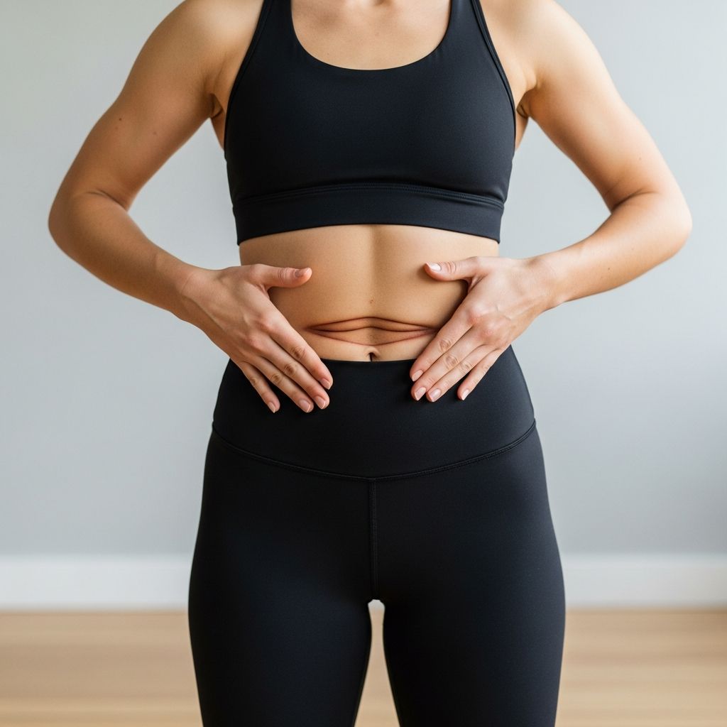
Diastasis recti, characterized by the separation of the rectus abdominis muscles along the midline, affects many individuals, particularly postpartum women, due to the stretching of the linea alba connective tissue. This condition arises from increased intra-abdominal pressure during pregnancy, obesity, or improper lifting techniques, leading to core weakness that impacts daily function. While often associated with a visible abdominal bulge, it can also contribute to back pain, pelvic floor issues, and postural imbalances. Effective recovery focuses on targeted rehabilitation rather than traditional crunches, emphasizing deep core activation and professional guidance for optimal healing.
Recognizing the Signs of Abdominal Muscle Separation
Early identification of diastasis recti is crucial for preventing long-term complications. A primary indicator is a soft, jelly-like bulge or ‘doming’ along the abdominal midline, especially noticeable when performing activities that increase intra-abdominal pressure, such as coughing or rising from a supine position. This doming occurs because the weakened linea alba fails to hold the muscles together under strain.
Other common symptoms include persistent lower back pain stemming from core instability, which compromises spinal support during movement. Individuals may struggle with posture, experiencing hip or pelvic discomfort as the body compensates for the lack of abdominal strength. Functional limitations often emerge, such as difficulty lifting objects, engaging in exercise, or even getting out of bed without discomfort. Pelvic floor dysfunction, manifesting as urinary incontinence or a sensation of pressure, frequently co-occurs due to the interconnectedness of core and pelvic muscles.
Not all cases present severe symptoms; some individuals notice only a subtle loss of core strength or a persistent ‘pooch’ above or below the belly button. Self-assessment involves lying on your back with knees bent, lifting the head slightly, and palpating the midline for a gap wider than two fingers, particularly around the navel. However, professional diagnosis by a physical therapist or physician is recommended to differentiate it from hernias and assess severity.
Risk Factors and Prevalence in Postpartum Women
Pregnancy is the leading risk factor for diastasis recti, as hormonal changes like relaxin soften connective tissues, while the growing uterus exerts sustained pressure on the abdominal wall. This separation often widens in the second and third trimesters and may persist postpartum if not addressed. Other contributors include multiple pregnancies, large babies, obesity, and labor strain.
Though more prevalent in women, men can develop it from heavy lifting, chronic coughing, or significant weight fluctuations. Studies indicate up to 60% of postpartum women experience some degree of separation at six weeks after delivery, with many resolving naturally within months, but 30-40% requiring intervention. Early screening during routine postpartum checkups can guide timely rehabilitation.
Building a Strong Foundation for Core Rehabilitation
Conservative management through physical therapy is the first-line treatment, promoting healing without surgery for most cases. Recovery begins with foundational exercises that activate the transverse abdominis (TA), the deepest abdominal layer acting like an internal corset. These movements prioritize gentle, controlled engagement over intensity to avoid exacerbating the separation.
A structured program typically spans phases: initial foundation building, progressive strengthening, and functional integration. Start with diaphragmatic breathing to coordinate breath with core activation, reducing intra-abdominal pressure. Progress to pelvic tilts and heel slides on the floor, focusing on drawing the belly button toward the spine without holding breath.
- Diaphragmatic Breathing: Lie supine, place hands on belly and chest. Inhale deeply through nose to expand belly (not chest), exhale slowly while gently contracting deep abs. Repeat 10 times daily.
- Pelvic Tilts: On back with knees bent, tilt pelvis to flatten lower back against floor, hold 5 seconds. 10-15 reps, 2-3 sets.
- Heel Slides: Supine, slide one heel away while maintaining core engagement, alternate legs. Build to 10 per side.
Manual therapy from a pelvic floor specialist can enhance tissue mobility, addressing tightness in related muscles. Real-time movement analysis corrects compensatory patterns, ensuring safe progression.
Safe Progression: Intermediate Exercises for Core Strength
Once foundational control is established (typically 4-6 weeks), introduce intermediate exercises to bridge the gap and narrow the linea alba. These target the TA, obliques, and pelvic floor synergistically, improving stability without traditional flexion like sit-ups, which can worsen doming.
- Bird-Dog: On hands and knees, extend opposite arm and leg while keeping spine neutral and core braced. Hold 5-10 seconds, 8-10 reps per side.
- Dead Bug: Supine, arms toward ceiling, lower opposite arm and leg slowly without arching back. 10 reps per side.
- Side-Lying Leg Lifts: Strengthen obliques by lifting top leg with bottom knee bent for stability. 10-12 reps per side.
Monitor for doming during exercises; if present, regress to easier variations. Consistency—20-30 minutes daily—yields results in 3-9 weeks per small studies. Combine with postural training to support spinal alignment.
Advanced Strategies and When to Escalate Treatment
For persistent gaps over 2-3 cm after 6 months of therapy, advanced options like electromagnetic muscle stimulation (e.g., Emsculpt) can supplement rehab by inducing supramaximal contractions. Surgery, such as abdominoplasty, is reserved for severe cases with hernias or skin laxity, involving muscle plication.
Lifestyle modifications aid recovery: maintain a healthy weight, avoid heavy lifting (>20 lbs initially), and use proper body mechanics. Supportive garments provide temporary stability but aren’t curative.
| Exercise Level | Focus | Samples | Reps/Sets |
|---|---|---|---|
| Beginner | Deep Core Activation | Breathing, Tilts | 10 reps, 2-3 sets |
| Intermediate | Stability | Bird-Dog, Dead Bug | 8-12 reps/side, 2 sets |
| Advanced | Integration | Planks (modified), Squats | 20-30 sec holds, 3 sets |
Common Mistakes That Worsen Abdominal Separation
Avoid exercises promoting forward flexion or twisting, like crunches, Russian twists, or full sit-ups, as they strain the linea alba. High-impact activities (running, jumping) before core readiness increase pressure. Over-reliance on surface abs neglects deep layers. Always exhale during exertion to manage pressure.
Frequently Asked Questions
Can diastasis recti heal without treatment?
Yes, mild cases often resolve naturally within 6-12 months postpartum, but therapy accelerates closure and prevents issues.
How long until I can do planks?
Typically 8-12 weeks with consistent rehab; start modified on knees.
Is surgery always needed?
No, only for severe, non-responsive cases; 90% improve with conservative care.
Can men get diastasis recti?
Yes, from obesity or heavy lifting.
When should I see a specialist?
If gap >2 fingers persists after 8 weeks or symptoms worsen.
References
- Understanding Diastasis Recti: Causes, Symptoms, and Effective Treatments — Eternal Wellness Medical Spa. 2023. https://eternalwellnessmedicalspa.com/blog/diastasis-recti/
- Diastasis Recti (DR): Causes, Symptoms, and Treatment — Mendwell Pelvic Health. 2024. https://www.mendwellhealth.com/all-conditions/diastasis-recti
- Diastasis Recti Rehabilitation — NCBI Bookshelf, StatPearls. 2023-10-25. https://www.ncbi.nlm.nih.gov/books/NBK573063/
- Understanding Diastasis Recti: Symptoms & Treatment — Look Natural. 2023. https://www.looknatural.com/blog/diastasis-recti-what-you-need-to-know
- How to Treat Diastasis Recti with Treatments and Symptoms — Henry Mayo Newswise. 2021-06-01. https://www.henrymayo.com/news-publications/news/2021/how-to-treat-diastasis-recti/