Understanding Brain Imaging in Newborns and Infants
When a newborn arrives in the world, parents naturally want to ensure their child is developing properly. One of the most valuable diagnostic tools available to pediatricians and neonatologists is cranial ultrasound, a sophisticated yet gentle imaging technique that allows medical professionals to visualize the developing brain without exposing infants to harmful radiation. This non-invasive procedure has become standard practice in neonatal intensive care units (NICUs) and pediatric hospitals worldwide, providing crucial insights into brain structure and function during the most critical period of neurological development.
The Science Behind Sound Wave Brain Imaging
Cranial ultrasound operates on a straightforward yet elegant principle: sound waves travel through body tissues at different speeds depending on tissue density, creating echoes that are converted into visual images displayed on a monitor. Unlike computed tomography (CT) scans or magnetic resonance imaging (MRI), which require the use of ionizing radiation or strong magnetic fields, ultrasound relies entirely on acoustic energy, making it exceptionally safe for use with vulnerable newborn patients.
The fundamental advantage of using ultrasound for infant brain imaging lies in the anatomical architecture of newborns. During infancy, particularly in the first months of life, the skull bones have not yet fully calcified or fused together. These incomplete skeletal structures create natural openings called fontanelles—commonly known as soft spots—which serve as acoustic windows through which ultrasound waves can freely penetrate to reach the brain tissue beneath. Once a child’s skull bones fully ossify and close these gaps, typically between 12 and 24 months of age, traditional ultrasound examination of the brain becomes impossible, as bone effectively blocks the passage of sound waves.
Key Anatomical Windows for Brain Examination
Sonographers and radiologists utilize several anatomical pathways to obtain comprehensive views of the infant brain:
- Anterior fontanelle: The primary acoustic window, located where the frontal and parietal bones meet at the top front of the infant’s head. This opening provides access to most brain structures and is used for the majority of imaging.
- Posterior fontanelle: Located at the back of the head where parietal and occipital bones meet, this smaller opening supplements imaging of posterior brain regions.
- Mastoid fontanelle: Positioned behind the ear, this window allows evaluation of lateral brain structures and blood flow patterns.
- Temporal region: The area near the temple provides additional access for specialized Doppler studies examining cerebral blood circulation.
Medical Conditions Detected Through Cranial Ultrasound
Pediatricians order cranial ultrasounds for multiple clinical scenarios. In premature infants, the procedure serves as routine screening to identify complications associated with early birth. The developing brain of a preterm infant faces specific risks including intraventricular hemorrhage (bleeding within the brain ventricles), periventricular leukomalacia (tissue damage in the white matter), and hypoxic-ischemic encephalopathy resulting from insufficient oxygen and blood flow during delivery.
Beyond prematurity-related concerns, cranial ultrasound helps physicians evaluate:
- Suspected structural brain malformations or congenital abnormalities
- Abnormal head size or shape, including microcephaly or macrocephaly
- Seizures or other neurological symptoms that suggest central nervous system dysfunction
- Suspected head trauma or birth injuries
- Brain infections acquired during pregnancy or after birth
- Hydrocephalus, characterized by excessive cerebrospinal fluid accumulation within brain ventricles
- Craniosynostosis, a condition where skull sutures prematurely fuse
- Signs of intrauterine growth restriction or genetic syndromes affecting neurological development
The Examination Procedure: What Parents Should Expect
Preparation for a cranial ultrasound examination is minimal and non-invasive. Unlike many medical procedures requiring fasting or medication administration, a head ultrasound demands no special physical preparation. Medical staff may suggest having a pacifier or bottle available to comfort and soothe the infant during the examination, as a calm baby produces clearer images.
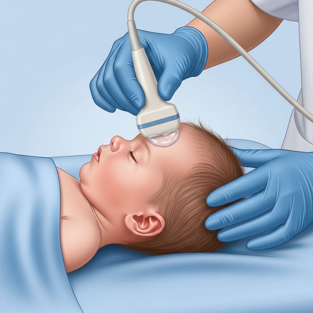
The actual procedure typically occurs at the infant’s bedside, whether in the NICU or an ultrasound suite. The sonographer begins by positioning the baby in a supine (face-up) position for optimal access to the anterior fontanelle. A clear, water-based acoustic gel is then applied to the ultrasound transducer—the handheld probe that emits and receives sound waves. This gel serves a critical function: it eliminates air pockets between the transducer and skin surface, allowing sound waves to travel unimpeded into the brain tissue. The sonographer gently applies the transducer to the soft spot, adjusting the angle and position to capture multiple imaging planes and perspectives of the brain structures.
Throughout the examination, real-time images appear immediately on a video monitor, allowing the sonographer to visualize the brain tissue and cerebrospinal fluid surrounding the brain and filling the brain’s ventricles. The entire imaging session typically requires 15 to 30 minutes, depending on the complexity of the clinical question and the number of structures requiring detailed assessment. Most infants tolerate the procedure well, as the gentle pressure and gel application cause no discomfort.
Advanced Imaging Techniques: Beyond Standard Views
Modern cranial ultrasound incorporates sophisticated Doppler technology that extends diagnostic capabilities beyond simple structural visualization. Doppler ultrasound measures the velocity and direction of blood flow through cerebral arteries, providing functional information about brain perfusion and circulation. This technique proves particularly valuable in detecting conditions like hypoxic-ischemic encephalopathy, where altered blood flow patterns can indicate brain injury severity and help predict long-term neurological outcomes.
Sonographers performing Doppler evaluation typically examine the anterior cerebral artery and middle cerebral artery, calculating a resistance index that reflects vascular properties and tissue perfusion. By comparing resistance measurements obtained at different times after birth—typically between 48 and 72 hours for encephalopathy assessment—clinicians can track the evolution of brain injury and guide treatment decisions.
Technical Parameters and Equipment Selection
The quality of cranial ultrasound images depends significantly on appropriate transducer selection and frequency optimization. Neonatal imaging typically employs high-frequency transducers ranging from 7.5 to 18 megahertz (MHz), with specific frequencies selected based on the infant’s age and the brain region under examination.
For newborns, sonographers generally begin with a curvilinear transducer of approximately 11 MHz to obtain broad survey images of the entire brain, including the cerebrum, brainstem, and cerebellum. As infants grow and fontanelles progressively narrow and become deeper, lower frequencies (8 MHz) penetrate tissue more effectively. Linear array transducers operating at higher frequencies (14 to 18 MHz) provide superior resolution of superficial brain structures, including the cortex and extra-axial space, though at the cost of reduced penetration depth. Advanced protocols often employ both sector (curvilinear) and linear transducers, switching between them to optimize visualization of different anatomical layers and structures.
Imaging Planes and Comprehensive Brain Assessment
Thorough cranial ultrasound examination requires visualization in multiple imaging planes—essentially viewing the brain from different angles to ensure no pathology is missed. Coronal imaging, which views the brain from front to back as if slicing perpendicular to the spine, reveals structures at different depths within the head. Sagittal imaging, obtained from side-to-side perspectives, shows brain anatomy along the body’s midline and slightly off-center regions.
During a complete examination, the sonographer assesses multiple anatomical parameters including normal brain anatomy recognition, evidence of brain maturation appropriate for the infant’s gestational age, the echogenicity (brightness) of brain tissue (parenchyma), ventricular diameter measurements, vessel patency and flow characteristics, and evaluation of extra-axial structures and fluid spaces. This systematic approach ensures consistent, reproducible examinations that facilitate tracking changes over time when serial ultrasounds are performed to monitor evolving conditions.
Safety Profile and Radiation Considerations
One of the most compelling advantages of cranial ultrasound in neonatal care is its exceptional safety profile. Unlike radiographic procedures or CT scanning, ultrasound imaging does not employ ionizing radiation, which carries risks including cellular DNA damage and increased long-term cancer risk, particularly concerning in the developing brain. Additionally, ultrasound examination requires no sedation or anesthesia, eliminating medication-related risks in vulnerable newborns. The procedure is painless, causes no tissue heating or damage, and can be repeated as many times as clinically necessary without safety concerns.
These characteristics establish cranial ultrasound as an ideal initial imaging modality for brain evaluation in infants, serving as a reliable screening tool complementary to more advanced imaging techniques like CT or MRI when those studies become necessary.
Portability and Accessibility in Neonatal Care
The technical simplicity and minimal equipment requirements of cranial ultrasound make it uniquely suited to the neonatal intensive care environment. Unlike CT or MRI examinations, which require transporting critically ill infants to imaging departments—often necessitating disconnection from life support equipment and monitoring systems—cranial ultrasound can be performed at the infant’s bedside without any repositioning or transport. A portable ultrasound machine and transducer represent the only equipment necessary, and examinations can be completed in minutes rather than the 30 to 60 minutes typically required for advanced imaging modalities.
This bedside capability proves invaluable for acutely ill premature infants in the NICU who may be too unstable for transport or for whom any intervention carries significant physiological stress. The speed, safety, and accessibility of cranial ultrasound have established it as the most widely used neuroimaging procedure in premature infants worldwide.
Interpreting Results and Clinical Significance
Normal cranial ultrasound findings demonstrate symmetrical brain structures, appropriately sized ventricles, smooth cortical surfaces consistent with the infant’s gestational age, and no evidence of hemorrhage, infection, or structural malformations. The cerebrospinal fluid surrounding the brain appears uniformly dark (hypoechoic), while the cortex demonstrates expected echogenicity appropriate for developmental stage.
Abnormal findings range from minor variants of no clinical significance to serious pathology requiring immediate intervention. Intraventricular hemorrhage, classified into four grades based on severity and location, may resolve spontaneously or progress to hydrocephalus requiring drainage procedures. Areas of increased echogenicity (brightness) in the white matter may indicate periventricular leukomalacia, suggesting hypoxic injury with significant implications for future neurodevelopmental outcomes. Ventricular enlargement may reflect various pathological processes, from simply delayed cerebrospinal fluid resorption to obstructive hydrocephalus requiring surgical intervention.
Monitoring Brain Development Over Time
Serial cranial ultrasound examinations performed at specific intervals—often at birth, day three to five, day 10, and week four in high-risk infants—create a developmental record showing brain maturation progression. Premature infants demonstrate measurable changes in sulcal development, tissue echogenicity, and ventricular dimensions as the brain matures, and ultrasound effectively documents these expected changes or identifies deviations suggesting abnormal development. This monitoring function helps clinicians distinguish normal maturation from pathological processes and guides decision-making regarding treatment interventions and family counseling regarding long-term prognosis.
Frequently Asked Questions
Q: At what age can infants no longer have cranial ultrasound performed?
A: Once the fontanelles close and skull bones fully fuse—typically between 12 and 24 months of age—ultrasound cannot visualize the brain because bone blocks sound wave penetration. After this age, advanced imaging like MRI or CT becomes necessary for brain evaluation.
Q: Is cranial ultrasound safe to repeat multiple times?
A: Yes. Cranial ultrasound involves no ionizing radiation, no sedation requirements, and no known harmful effects. It can be safely repeated as many times as clinically necessary to monitor brain conditions or track development.
Q: Can cranial ultrasound definitively rule out all brain abnormalities?
A: While cranial ultrasound effectively detects many significant abnormalities, some subtle structural variations or certain malformations may not be apparent on ultrasound. MRI provides superior detail for some conditions and may be recommended if clinical suspicion remains high despite normal ultrasound findings.
Q: How long does a cranial ultrasound examination typically take?
A: Standard cranial ultrasound examinations usually require 15 to 30 minutes depending on the clinical question, the infant’s cooperation, and the number of specific structures requiring detailed assessment.
Q: Does the baby need to be completely still during the ultrasound?
A: While minimal movement is ideal for image quality, some infant movement is expected and acceptable. A pacifier, bottle, or gentle holding by a parent can help keep the baby calm and still, improving imaging quality.
References
- Cranial (Head) Ultrasound — Radiologyinfo.org. 2024. https://www.radiologyinfo.org/en/info/ultrasound-cranial
- Cranial ultrasound for beginners — Caro-Domínguez P, et al. PubMed Central/NIH. 2021. https://pmc.ncbi.nlm.nih.gov/articles/PMC8107866/
- Pediatric Ultrasound — Ann & Robert H. Lurie Children’s Hospital of Chicago. 2024. https://www.luriechildrens.org/en/specialties-conditions/ultrasound/
- Neonatal Cranial ultrasound — British Medical Ultrasound Society. 2024. https://www.bmus.org/media/resources/Susannah_Lightfoot_Neonatal_Cranial_ultrasound.pdf
- Pediatric Head Ultrasounds — Cleveland Clinic. 2024. https://www.youtube.com/watch?v=E5pLJxMmU-8
- Neonatal Brain US — Radiology Assistant. 2024. https://radiologyassistant.nl/pediatrics/spine/neonatal-brain-us