Understanding Ear Deformities: When the Helical Rim Doesn’t Develop Normally
Ear deformities present at birth are among the most common congenital conditions pediatricians encounter. One particular category of these deformities—constricted ears—affects the structural integrity of the upper portion of the ear. Understanding this condition is essential for parents, as early recognition and intervention can lead to significantly better outcomes for their children.
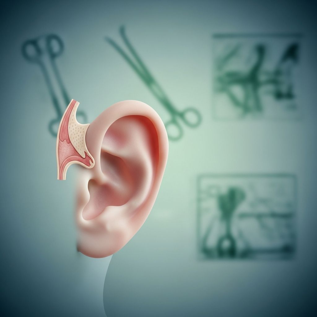
Constricted ears, also referred to as lop ears or cup ears in medical literature, occur when the helical rim (the outer curved edge of the ear) develops abnormally during fetal growth. Rather than maintaining a natural curved shape, the affected ear develops with a folded, wrinkled, or excessively tight appearance. This condition can manifest in varying degrees of severity, ranging from subtle cosmetic concerns to more pronounced deformities that may impact both appearance and function.
How Constricted Ears Form: The Development Story
During pregnancy, the external ear structures undergo a complex developmental process. The ear begins forming around the third to sixth month of gestation, during which time the cartilage and skin must develop in proper proportion and alignment. When this developmental process encounters disruptions, constricted ears can result.
The fundamental characteristic of constricted ears involves an insufficiency of skin or cartilage—or sometimes both—in the regions that comprise the helical rim and the scapha (the curved depression beneath the upper ear rim). This shortage forces the available tissue into patterns that create the characteristic folded or tight appearance. In milder presentations, only the uppermost portion of the helix may fold inward. In more severe cases, the cartilage itself becomes tightly rolled, substantially altering the ear’s normal contour.
Identifying the Root Causes: What Medical Science Currently Understands
The exact etiology of constricted ears remains incompletely understood by the medical community. However, research has identified several factors that may contribute to their development:
- Intrauterine positioning: The position of the developing fetus within the uterus may influence ear formation. Compression against the uterine wall or positioning that restricts blood flow to the developing ear can result in abnormal cartilage development.
- Reduced blood supply: Insufficient vascular supply to the outer ear during critical developmental windows can impair the formation of normal ear structures.
- Medication exposure: Maternal use of certain medications during pregnancy, particularly isotretinoin (commonly prescribed for severe acne) and thalidomide, has been associated with increased incidence of ear deformities.
- Prenatal infections: Viral infections such as rubella or influenza during specific pregnancy trimesters may increase the risk of congenital ear abnormalities.
- Genetic factors: While most cases of constricted ears occur sporadically without clear hereditary patterns, genetic mutations or inherited conditions can occasionally contribute to their development.
- Muscular development variations: Underdevelopment or atypical attachment of the superior auricular muscles can create tension that pulls ear structures into abnormal positions.
Importantly, research indicates that constricted ears develop randomly in many cases, and parents should understand that this condition typically does not result from anything they did or did not do during pregnancy.
Distinguishing Constricted Ears from Other Ear Conditions
Several related but distinct ear deformities exist in the broader category of congenital ear abnormalities. Understanding these distinctions helps parents and caregivers better comprehend their child’s specific condition:
| Condition Type | Key Characteristics | Primary Distinction |
|---|---|---|
| Constricted/Cup Ears | Helical rim is folded, wrinkled, or tight; involves shortage of skin or cartilage | Affects the upper rim structure specifically |
| Cryptotia | Upper portion of ear buried beneath scalp skin; appears partially hidden | Ear is covered by skin rather than fully protruding |
| Prominent Ears | Ears extend more than 2 centimeters from the head; underdeveloped antihelix | Normal structure but abnormal positioning and spacing |
| Stahl’s Ear | Extra fold of cartilage creates pointed appearance | Additional cartilage rather than insufficient tissue |
Clinical Presentation: How Parents Recognize Constricted Ears
Constricted ears are typically observable at birth or within the immediate newborn period, making early detection straightforward through routine pediatric examination. Parents may notice several characteristic features:
- The affected ear appears noticeably smaller than the opposite ear
- The upper portion of the ear is folded forward or appears compressed
- The ear rim may appear wrinkled or corrugated rather than smooth
- In severe cases, the ear may protrude abnormally from the side of the head due to malformed cartilage
- The deformity may affect one ear or both ears
- The severity can range from subtle to dramatic, depending on the degree of cartilage and skin deficiency
While the cosmetic appearance is the most obvious feature parents notice, it is important to note that constricted ears may occasionally be associated with other developmental considerations. Some children may experience ear canal abnormalities or susceptibility to recurrent ear infections, though hearing development itself typically proceeds normally.
Early Intervention: The Critical Window for Non-Surgical Treatment
One of the most important advances in managing constricted ears involves early non-surgical intervention through ear molding. This approach takes advantage of the remarkable plasticity of infant cartilage during the neonatal period.
Ear molding works on a fundamental principle: newborn ear cartilage remains exceptionally soft and malleable due to residual maternal hormones and the cartilage’s inherent lack of full calcification. By applying gentle, consistent pressure through specially designed molding devices during this window of opportunity—ideally within the first two weeks of life—the ear can be gradually reshaped into a more normal configuration.
The molding process involves placing a soft plastic splint beneath the helix of the upper ear. This splint is carefully positioned and maintained by trained specialists who understand both the biomechanics of ear cartilage and the specific needs of neonatal patients. As the infant grows and develops, new tissue forms in response to the sustained but gentle pressure from the mold, gradually establishing a more normal ear shape.
The success of ear molding is significantly higher when initiated early. Studies have demonstrated that treatment beginning within the first two to three weeks of life achieves optimal results, as the cartilage responds most readily to molding pressure during this period. Waiting beyond six to eight weeks substantially decreases the likelihood of successful non-surgical correction.
Surgical Options for More Severe Cases
In situations where constricted ears are more severe, non-surgical molding proves insufficient, or when ear molding was not performed during infancy, surgical reconstruction becomes necessary. Otoplasty—the surgical reshaping of the ear—offers a definitive solution for correcting these deformities.
Surgical intervention typically occurs after the ear has reached approximately 85-90% of its adult size, which generally occurs around five to six years of age. At this point, the ear is sufficiently developed to allow for surgical planning and execution while still being young enough that the child benefits from correction during their formative years.
The surgical approach varies depending on the specific nature of the deformity. Reconstructive surgery may involve:
- Repositioning and reshaping of existing cartilage to restore normal helical contour
- Creating new cartilage structures where tissue is absent, sometimes using cartilage grafts
- Adjusting skin coverage to accommodate the reshaped cartilage framework
- In severe cases where substantial ear structures are missing, more complex reconstruction may be required
Modern otoplasty techniques have evolved significantly, with surgeons employing sophisticated approaches that preserve ear function while achieving superior cosmetic outcomes. Success rates for surgical correction of constricted ears are quite high, with most children achieving satisfactory ear appearance and normal function.
Comprehensive Monitoring and Development Support
Regardless of whether a child receives non-surgical molding, surgical correction, or both, comprehensive monitoring throughout early childhood remains essential. Specialized pediatric teams track multiple developmental domains:
- Hearing assessment: Regular audiological evaluations ensure normal hearing development, even though constricted ears typically do not affect hearing
- Speech and language development: Monitoring ensures language acquisition proceeds normally
- Otologic health: Assessment for ear canal involvement or infection susceptibility
- Psychological adjustment: As the child grows, support for emotional adjustment regarding appearance is important
- Surgical healing and outcome: If surgery was performed, continued follow-up ensures optimal long-term results
Parental Guidance and Expectations
Parents of children with constricted ears benefit from understanding realistic timelines and expectations. If ear molding is initiated in infancy, visible improvements typically appear within weeks to a few months. Complete reshaping may take several months of consistent use of the molding device.
For families pursuing surgical correction, the timeline extends longer but offers definitive results. The surgical procedure itself is relatively brief, though recovery involves several weeks of healing before final results become apparent. Scars from otoplasty are typically minimal and hidden in the natural creases and contours of the ear.
Modern treatment approaches emphasize early intervention, whether through molding or eventual surgery, as addressing ear deformities during childhood years supports both functional development and psychological well-being during the crucial formative years.
Frequently Asked Questions
Will constricted ears affect my child’s hearing?
Constricted ears typically do not affect hearing development. While the external ear appearance is altered, the internal ear structures and hearing mechanisms usually develop normally. However, regular hearing assessments are recommended as part of comprehensive monitoring.
What is the ideal age to begin treatment?
For non-surgical ear molding, the ideal timing is within the first two weeks of life, with good results possible up to about eight weeks of age. For surgical correction, the typical recommendation is between five and six years old, once the ear has reached approximately 85-90% of adult size.
Is ear molding painful for my baby?
Ear molding devices are designed to be soft and comfortable. The procedure does not cause pain, though some infants may need adjustment time. Trained specialists ensure the devices are properly fitted and comfortable.
Can constricted ears run in families?
While most cases of constricted ears occur sporadically without clear hereditary patterns, genetic factors can occasionally play a role. If you have family members with ear deformities, discuss this with your pediatrician.
What are the success rates for surgical correction?
Success rates for otoplasty and surgical ear reconstruction are quite high, with most children achieving satisfactory cosmetic and functional outcomes. Specific rates depend on the severity of the initial deformity and the surgical technique employed.
Will my child need multiple surgeries?
Most children with constricted ears require only one surgical procedure if they undergo otoplasty. However, in cases involving more severe deformities with missing ear structures, staged surgical reconstruction may be necessary.
References
- Prominent or Constrained Ears — Atrium Health Wake Forest Baptist. 2024. https://www.wakehealth.edu/condition/p/prominent-or-constricted-ears
- Constricted/Lop/Cup Ears — Children’s Hospital of Philadelphia. 2024. https://www.chop.edu/conditions-diseases/constricted-ears
- Constricted Ears: A Guide to Diagnosis and Treatment — EarWell Centers. 2024. https://earwellcenters.com/constricted-ears/
- Congenital Deformities of the Outer Ear — Yale Medicine. 2024. https://www.yalemedicine.org/conditions/congenital-deformities-of-the-outer-ear
- Pediatric Ear Malformations – Conditions and Treatments — Children’s National Hospital. 2024. https://www.childrensnational.org/get-care/health-library/ear-malformations
- Constricted Ear — St. Louis Children’s Hospital. 2024. https://www.stlouischildrens.org/conditions-treatments/constricted-ear
- Cryptotia Ear Deformity: The Path from Diagnosis to Treatment — EarWells. 2024. https://www.earwells.com/our-blog/cryptotia-ear/
- Ear Deformities: Types, Causes & Treatment — Cleveland Clinic. 2024. https://my.clevelandclinic.org/health/diseases/24904-ear-deformity
- Constricted Ear — Boston Children’s Hospital. 2024. https://www.childrenshospital.org/conditions-treatments/constricted-ear