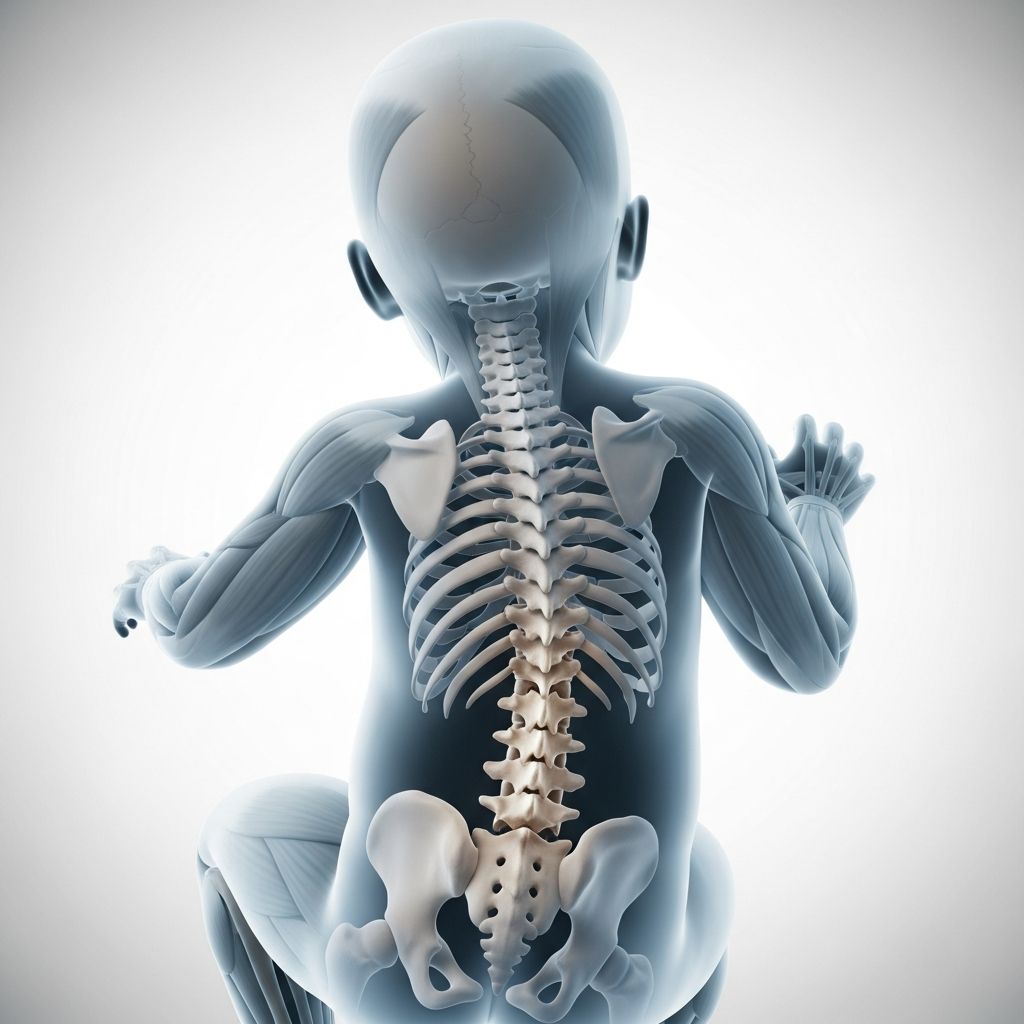
Congenital scoliosis is a rare spinal deformity present at birth, characterized by a sideways curvature of the spine due to malformed vertebrae formed during fetal development. Affecting approximately 1 in 10,000 newborns, it differs from more common adolescent idiopathic scoliosis by originating from structural defects in the spine’s bony segments.
Understanding the Spinal Development Process
The human spine begins forming in the early weeks of pregnancy, specifically between the 4th and 8th weeks of gestation when vertebrae differentiate from a single tissue column. Disruptions during this critical embryological phase can lead to anomalies that manifest as congenital scoliosis. These defects include incomplete vertebral formation or failure of vertebrae to separate properly, resulting in an asymmetrical spine that curves laterally, often in a C or S shape.
Normally, each vertebra develops symmetrically, but in congenital cases, one side may grow differently, pulling the spine into a curve as surrounding muscles and ligaments exert uneven forces. This rigidity distinguishes it from flexible curves seen in other scoliosis types, making progression harder to predict but often necessitating close monitoring.
Primary Causes of Vertebral Anomalies
Congenital scoliosis arises from two main categories of vertebral malformations:
- Failure of Formation: Parts of one or more vertebrae do not fully develop, creating wedge-shaped or hemivertebrae (half-formed vertebrae). This instability leads to a sharp spinal angle that may worsen with growth.
- Failure of Segmentation: Adjacent vertebrae fail to separate, forming a bony bar that restricts growth on one side. This unilateral tethering progressively curves the spine as the child grows.
Combinations of these defects, such as a bony bar on one side and a hemivertebra on the opposite, pose the greatest risk for rapid progression and severe deformity. The exact triggers remain unclear, potentially involving genetic or environmental factors during early pregnancy, though it is generally non-hereditary.
Recognizing Early Signs in Newborns
Many cases are spotted during routine newborn exams due to visible back asymmetries, such as a prominent rib hump or uneven shoulder levels. However, milder curves might evade detection until later infancy or even adolescence, when clothing fits poorly or a bathing suit reveals the issue.
Symptoms are typically absent in early stages since the condition is painless initially. As curves advance, potential indicators include:
- Uneven hips or waistline
- One shoulder blade protruding more than the other
- Limited spinal flexibility
- In severe instances, breathing difficulties from rib cage distortion or neurological issues like weakness if the spinal cord is compressed
Associated conditions heighten concern: up to 50-70% of cases link to anomalies in the heart, kidneys, urinary tract, or spinal cord (e.g., tethered cord syndrome), underscoring the need for systemic screening.
Diagnostic Approaches for Accurate Assessment
Diagnosis starts with a physical exam using the Adams forward bend test, where the child bends forward to reveal trunk asymmetry. Confirmation relies on imaging:
| Method | Purpose | Details |
|---|---|---|
| X-rays | Measure curve magnitude (Cobb angle >10° confirms scoliosis) | Full spine views assess curve location, rigidity, and vertebral defects |
| MRI | Evaluate spinal cord/nerve involvement | Detects hidden issues like syringomyelia or dysraphism |
| CT Scans | Detailed bone anomaly visualization | Used sparingly in infants due to radiation; aids surgical planning |
| Ultrasound/Echo | Screen associated organ defects | Checks kidneys, heart; renal ultrasounds routine |
Regular follow-ups track progression, as 10-25% of mild curves stabilize, while others advance unpredictably.
Monitoring and Observation Strategies
For curves under 20-25° without aggressive growth imbalance, observation suffices. Parents track changes at home, while orthopedists schedule X-rays every 4-6 months in infancy, spacing out as stability emerges. Factors favoring watchful waiting include balanced anomalies (e.g., opposing hemivertebrae) and no neurological risks.
Non-Surgical Treatment Options
Bracing aims to guide growth in flexible curves (20-40°), typically using custom orthoses worn 16-23 hours daily until skeletal maturity. Effectiveness varies; rigid congenital curves respond less predictably than idiopathic ones.
Serial casting, a plaster-based method, temporarily corrects curves in very young children (under 3), changed every 2-3 months under anesthesia. It buys time before definitive intervention.
Surgical Interventions for Progression Control
Surgery becomes essential for curves exceeding 40-50°, rapid progression (>5°/year), or neurological compromise. Goals: halt worsening, balance growth, preserve function.
Common procedures:
- Growing Rods: Dual rods attached to spine anchors, lengthened surgically every 6 months to match growth. Removed post-puberty for fusion.
- Vertical Expandable Prosthetic Titanium Rib (VEPTR): Supports chest wall and spine in young patients, expandable non-surgically.
- Spinal Fusion: Joins vertebrae with bone grafts/rods; deferred until age 5+ for adequate fusion bed.
- Hemivertebra Excision: Removes malformed segments, realigns spine; ideal for isolated defects.
Timing weighs risks: early surgery prevents deformity but demands multiple operations; delaying risks pulmonary compromise.
Potential Complications and Associated Risks
Beyond spinal progression, children face:
- Cardiac/renal malformations requiring specialist care
- Respiratory issues from thoracic distortion
- Neurological deficits (rare, 10-20%)
- Psychosocial impacts from bracing/surgery
Long-Term Prognosis and Family Support
Outcomes hinge on defect severity and timely intervention. Mild cases often need no treatment, yielding normal adulthood. Severe ones may require lifelong monitoring, but modern techniques yield 80-90% curve control and good function.
Families benefit from multidisciplinary teams (orthopedists, cardiologists, nephrologists) and support groups for emotional resilience.
Frequently Asked Questions (FAQs)
What is the likelihood of my child developing congenital scoliosis?
It affects 1 in 10,000 newborns, far rarer than adolescent scoliosis.
Is congenital scoliosis hereditary?
Most cases are sporadic, not directly inherited, though genetic factors may contribute.
Can congenital scoliosis be prevented?
No, as causes are embryological insults beyond control, but prenatal care aids overall health.
When is surgery typically recommended?
For progressive curves >40°, imbalance, or symptoms, often after failed bracing.
Does congenital scoliosis cause pain in babies?
Rarely in infancy; discomfort arises later with severe curves.
How often are check-ups needed?
Every 4-6 months initially, based on growth velocity.
References
- Congenital Scoliosis — Columbia Orthopedic Surgery. 2023. https://www.columbiaortho.org/patient-care/specialties/pediatric-orthopedics/conditions-treatments/spine-disorders-scoliosis/congenital-scoliosis
- Congenital Scoliosis — Children’s Hospital of Philadelphia (CHOP). 2024-05-15. https://www.chop.edu/conditions-diseases/congenital-scoliosis
- Congenital Scoliosis — National Center for Biotechnology Information (PMC), NIH. 2012-10-01. https://pmc.ncbi.nlm.nih.gov/articles/PMC3468011/
- Congenital Scoliosis — Parkland Health. 2023. https://www.parklandhealth.org/congenital-scoliosis
- Congenital Scoliosis: What It Is, Symptoms & Treatment — Cleveland Clinic. 2024-02-20. https://my.clevelandclinic.org/health/diseases/congenital-scoliosis
- Congenital Scoliosis — American Academy of Orthopaedic Surgeons (AAOS) OrthoInfo. 2023-06-01. https://orthoinfo.aaos.org/en/diseases–conditions/congenital-scoliosis/