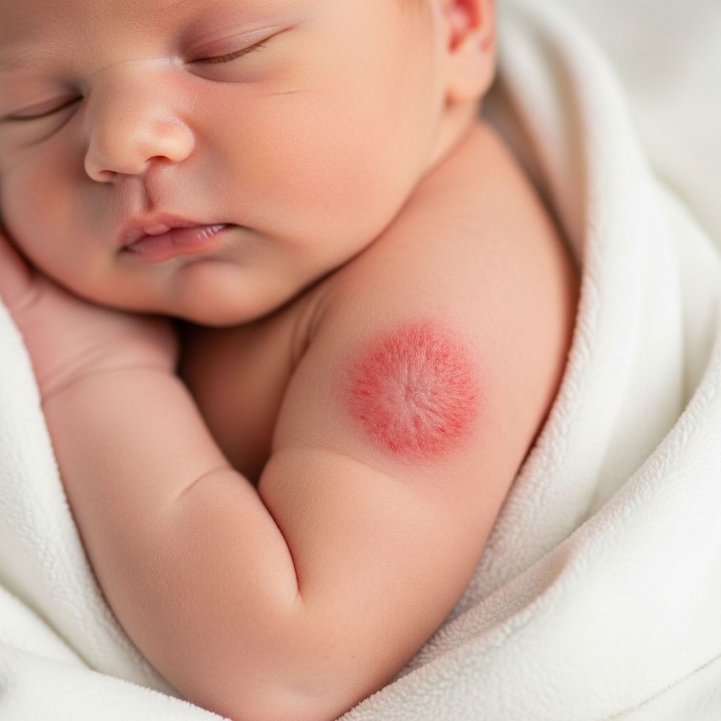
Congenital hemangiomas are rare vascular birthmarks formed by clusters of extra blood vessels that develop fully in the womb and are visible at birth, distinguishing them from more common infantile hemangiomas that grow postnatally.
Defining Congenital Vascular Birthmarks
These growths arise from abnormal proliferation of blood vessel cells during fetal development, resulting in a fully mature lesion by birth. Unlike infantile hemangiomas, which expand after birth and often regress spontaneously, congenital variants reach maximum size prenatally and exhibit varied behaviors. They typically manifest as solitary lesions, though multiple occurrences are possible, and affect approximately 1-2% of newborns, far less frequently than infantile types.
Key characteristics include a well-defined border, warm texture due to superficial vessels, and colors ranging from pink to deep purple. Locations favor extremities, head, and neck, though trunk involvement occurs. Parents often notice them immediately post-delivery, prompting early medical evaluation.
Primary Categories of Congenital Hemangiomas
Congenital hemangiomas classify into three main subtypes based on their natural progression: rapidly involuting (RICH), non-involuting (NICH), and partially involuting (PICH). Each type dictates monitoring and intervention strategies.
- RICH (Rapidly Involuting Congenital Hemangioma): These shrink dramatically within months, often resolving by 12-18 months with minimal residue. Initial presentation includes a raised, purple plaque that ulcerates or fades progressively.
- NICH (Non-Involuting Congenital Hemangioma): Persistent lesions maintain size and shape lifelong, appearing as firm, oval plaques with coarse surface vessels. They rarely regress without intervention.
- PICH (Partially Involuting Congenital Hemangioma): A hybrid form where portions regress while others persist, leading to irregular involution patterns observable over 1-2 years.
| Type | Behavior | Timeline | Common Residue |
|---|---|---|---|
| RICH | Rapid shrinkage | 12-18 months | Atrophic skin, veins |
| NICH | No involution | Lifelong stable | Persistent plaque |
| PICH | Partial regression | Variable, 1-2 years | Mixed atrophic/persistent |
Recognizing Early Indicators in Infants
At birth, lesions appear as solitary, round or oval patches measuring 1-10 cm, with hues from erythematous pink to violaceous blue. Superficial subtypes show waxy, telangiectatic surfaces; deeper ones feel spongy and warm. A peripheral pale halo may encircle the growth, and palpation reveals pulsatility or bruit in vascular-rich cases.
Complications, though uncommon, include ulceration (especially in RICH), bleeding, or consumptive coagulopathy (Kasabach-Merritt phenomenon, rare in congenital types). Functional impairments arise if near eyes, mouth, or airways. Head/neck sites heighten cosmetic and functional concerns.
Diagnostic Approaches for Accuracy
Diagnosis relies on clinical exam, but imaging confirms extent. Prenatal ultrasound detects large head/neck lesions as hypoechoic masses with vascular flow. Postnatally, Doppler ultrasound assesses flow patterns—high-velocity arteries in RICH/NICH differentiate from infantile hemangiomas.
MRI or angiography evaluates deep involvement or complications. Biopsy is rarely needed but reveals lobular capillary architecture without GLUT1 positivity, contrasting infantile hemangiomas. Specialist referral to pediatric dermatology or vascular anomaly centers ensures precise subtyping.
Observation and Monitoring Protocols
For asymptomatic RICH, active non-intervention suffices, with serial photos tracking involution. Monthly pediatric checks monitor for ulceration or infection. NICH/PICH demand baseline imaging and annual reviews for growth or symptoms.
- Photograph lesions monthly for progression logs.
- Assess vital signs; watch for heart strain in large lesions.
- Educate families on hygiene to prevent secondary issues.
Multidisciplinary teams—including dermatologists, surgeons, and psychologists—support families, addressing emotional impacts.
Medical Interventions for Persistent Cases
Pharmacotherapy is limited; beta-blockers like propranolol, effective for infantile hemangiomas, show inconsistent results in congenital types due to absent growth phase. Steroids or vincristine target complications like coagulopathy.
Pulsed dye laser treats surface telangiectasia, improving aesthetics without affecting bulk. For refractory NICH, sirolimus (mTOR inhibitor) shows promise in stabilizing growth per recent case series.
Surgical Options and Timing
Excision suits NICH/PICH post-involution (age 1-2 years) or urgent cases impairing function. RICH rarely requires surgery unless residual deformity persists. Preoperative embolization minimizes bleeding in vascular lesions.
Techniques include tangential shave for superficial sites or full-thickness resection with reconstruction. Outcomes favor early intervention for optimal cosmesis.
Potential Risks and Complication Management
High-output failure occurs in extensive lesions (>10% body surface), necessitating cardiology input. Ulceration risks infection; treat with barriers, antimicrobials. Rare heart overload or thrombocytopenia warrants hematology.
Long-term, atrophic scars or telangiectasia may need laser revision. Psychological support mitigates stigma, especially facial lesions.
Everyday Care Tips for Parents
Gentle cleansing with emollients prevents irritation. Avoid trauma; use protective clothing. Sun protection post-involution guards hypopigmented areas.
- Monitor for color/size changes weekly.
- Report fever, bleeding, or rapid expansion immediately.
- Join support networks for shared experiences.
Nutrition and routine immunizations proceed normally unless contraindicated.
Prognosis Across Subtypes
RICH boasts excellent prognosis, with 90% full resolution by age 2. NICH persists benignly but may prompt elective removal for aesthetics. PICH yields intermediate outcomes. Overall, malignancy risk is negligible.
Follow-up until school age ensures comprehensive resolution assessment.
Frequently Asked Questions
Can congenital hemangiomas be detected during pregnancy?
Yes, prenatal ultrasound may identify large lesions on head or limbs as vascular masses, guiding delivery planning.
Do all congenital hemangiomas require treatment?
No, many RICH regress without intervention; treatment targets complications or cosmetic issues in NICH/PICH.
Is surgery always successful for NICH?
Surgery effectively removes persistent NICH, but scarring varies by size/location; timing optimizes results.
Are congenital hemangiomas cancerous?
No, they are benign vascular tumors with no malignant potential.
How do they differ from strawberry hemangiomas?
Congenital types are birth-present and fully grown; infantile (strawberry) grow postnatally and often involute by age 5.
References
- Congenital haemangioma — Great Ormond Street Hospital. 2023. https://www.gosh.nhs.uk/conditions-and-treatments/conditions-we-treat/congenital-haemangioma/
- Non-Involuting Congenital Hemangioma (NICH) — Vascular Birthmarks Foundation. 2023. https://www.vbiny.org/conditions/vascular-tumors/non-involuting-congenital-hemangioma-nich/
- Congenital Hemangiomas — Nemours KidsHealth. 2023. https://kidshealth.org/en/parents/congenital-hemangioma.html
- Infantile Hemangioma — Children’s Hospital Colorado. 2023. https://www.childrenscolorado.org/conditions-and-advice/conditions-and-symptoms/conditions/infantile-hemangioma/
- Hemangiomas — Seattle Children’s Hospital. 2023. https://www.seattlechildrens.org/conditions/hemangiomas/