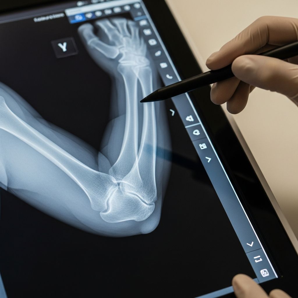
When a child hurts an arm, one of the first questions parents ask is whether the elbow might be broken. An elbow X-ray is the most common imaging test doctors use to check for fractures, dislocations, or other injuries in this joint.1 This guide explains in simple terms why an elbow X-ray might be ordered, how the test is done, what your child will experience, and how doctors interpret the images.
Why Doctors Order Elbow X-Rays for Kids
Pediatric elbows are particularly prone to injury because children are active and their bones are still growing.3 An X-ray helps doctors see bones and some joint details that can’t be evaluated by a physical exam alone.
Common reasons for an elbow X-ray
- Recent fall on an outstretched hand or directly onto the elbow
- Sports injuries, such as from gymnastics, skateboarding, or contact sports
- Visible deformity, such as an elbow that looks out of place or crooked
- Refusal to move the arm or severe pain when trying to bend or straighten the elbow
- Swelling, bruising, or warmth around the joint
- Concerns about a growth plate (physis) injury
- Follow-up after a known fracture to check healing or the position of a cast or pins
Injuries and conditions an elbow X-ray can show
On X-ray, doctors can look for a variety of problems that affect the bones and joint alignment.3
- Fractures (breaks) in the upper arm bone (humerus), forearm bones (radius and ulna), or small bony prominences around the elbow
- Dislocations where the bones are no longer aligned properly at the joint
- Growth plate injuries that can affect how the bone grows in the future
- Signs of previous injury, such as healing fractures or early deformity
- Joint effusion (fluid in the joint) that may be a clue to a hidden fracture
How an Elbow X-Ray Works
X-rays use a small, carefully controlled amount of ionizing radiation to create images of the inside of the body. Dense tissues like bone block more X-rays and appear white or light on the picture, while softer tissues appear darker.3
Basic steps of the imaging process
- Your child is positioned between an X-ray tube (which sends out X-rays) and a digital detector or plate.
- The machine emits a brief pulse of X-rays that passes through the elbow.
- The detector captures the pattern of X-rays that makes it through the elbow and converts it into a digital image.
- A radiologist (a doctor specializing in imaging) reviews the images and writes a report for the ordering provider.
Standard views of the pediatric elbow
For most elbow injuries, doctors request at least two different views to see the joint from more than one angle.13
| View name | How the arm is positioned | What it helps show |
|---|---|---|
| Front (AP) view | Elbow straight or as straight as pain allows, palm facing up | Overall alignment of the elbow, visible fractures in humerus, radius, and ulna |
| Side (lateral) view | Elbow bent about 90 degrees, thumb up | Subtle fractures, joint effusion, relationships between bones |
| Additional or oblique views | Arm angled slightly in or out, as needed | Better visualization of small fractures or specific parts of the joint |
Preparing Your Child for the Exam
Most children tolerate an elbow X-ray very well, especially when parents know what to expect and can walk them through the process calmly.
Before you arrive
- Explain that the test is like “taking a picture of the bones” and does not hurt.
- Let your child know that they may need to hold very still for a few seconds at a time.
- Dress them in loose, comfortable clothing that is easy to roll up or remove from the injured arm.
- Leave jewelry at home, especially bracelets or watches on the affected arm.
At the imaging center or hospital
- You will usually check in at radiology or an imaging department.
- A technologist (often called an X-ray tech) will confirm your child’s identity and ask brief questions about the injury.
- Parents are often allowed to stay in the room, wearing a lead apron for protection, to comfort and coach the child.
What Happens During the Elbow X-Ray
The exam itself is quick, often taking less than 10–15 minutes, with only a few seconds of actual exposure for each image.
Positioning and comfort
- Your child may sit on a chair, stand, or lie on a table, depending on age, size, and pain level.
- The technologist gently places the injured arm in one position at a time and supports it to minimize movement.
- Soft pads, sponges, or sandbags may be used to help hold the arm comfortably.
- If the elbow is very painful, the technologist will adjust the position to reduce discomfort as much as possible.
Staying still and breathing
- Your child will be asked to hold very still for each picture.
- For most elbow X-rays, they can breathe normally; they do not usually need to hold their breath.
- Each image is taken in a fraction of a second, and the technologist may quickly check the pictures before moving to the next position.
Radiation Safety and Children
Parents often worry about radiation exposure. Medical experts emphasize that diagnostic X-rays use the lowest dose that will give a clear image, and the benefits of accurate diagnosis generally far outweigh the small risk from radiation.3
Why X-rays are considered safe when medically needed
- The dose from a single elbow X-ray series is typically very low, much less than that from larger studies such as CT scans.
- Modern digital equipment and strict protocols help minimize exposure, especially in children.
- Protective shields or lead aprons may be used to cover areas of the body not being imaged when appropriate.
How pediatric radiology teams reduce exposure
- Using child-sized settings and equipment to avoid unnecessary radiation
- Limiting the number of images to those that are truly needed
- Avoiding repeat images by careful positioning and immobilization techniques
- Following national guidelines and quality standards for pediatric imaging3
How Radiologists Read a Child’s Elbow X-Ray
Interpreting a pediatric elbow X-ray is more complex than reading an adult image because much of the joint is still made of cartilage, which does not show on X-ray.35 Radiologists use specific landmarks and patterns to tell normal development from injury.
Growth and development of the elbow
- At birth, the elbow is mostly cartilage and cannot be fully evaluated with standard X-rays.3
- As children grow, different ossification centers (areas where bone forms) appear in a predictable order.
- These centers gradually enlarge and fuse, forming the mature bony structures seen in adults.5
Key things doctors look for on the images
- Overall alignment of the upper arm and forearm bones
- Continuity of the bone cortex (outer edge) to identify subtle cracks or breaks
- Joint spaces and whether the bones meet properly at the elbow
- Indirect signs of injury, such as a visible joint effusion that can indicate a hidden fracture
- Comparison to typical developmental patterns for the child’s age
Why subtle injuries can be missed without expertise
Because normal developmental changes can look unusual, pediatric elbow X-rays are a common source of diagnostic errors if not interpreted carefully.13 This is why radiologists and emergency physicians often use systematic checklists and, when needed, comparison with the other elbow.
After the X-Ray: Getting and Understanding Results
Once the images are taken, they are reviewed by a radiologist who sends a report to the doctor who ordered the test. In emergency settings, a brief initial interpretation may be given quickly so treatment decisions can be made.
Possible types of findings
- No fracture or dislocation – bones look normal, though soft-tissue injury may still be present.
- Simple fracture – a break that is aligned well and may be treated with a splint or cast.
- Displaced fracture – bone pieces are shifted or angled; may require reduction and sometimes surgery.4
- Growth plate injury – monitored closely because it can affect bone growth.
- Joint dislocation – bones are out of place and need to be repositioned.
Questions to ask your child’s doctor
- What did the X-ray show? Is there a fracture or dislocation?
- Does the injury involve a growth plate?
- What treatment is needed now (splint, cast, sling, surgery, or observation)?
- How long will it take to heal, and will we need repeat X-rays?
- Are there any activity restrictions, such as avoiding sports or playground equipment?
When More Imaging May Be Needed
Most elbow injuries in children can be evaluated adequately with X-rays.3 In some situations, however, additional tests may be recommended.
- Repeat X-rays a week or two later to see healing changes that make a subtle fracture more obvious.
- Ultrasound to assess soft tissues in very young children when X-rays are less clear.
- MRI to evaluate cartilage, ligaments, or complex injuries not visible on X-ray.
- CT scan in select cases where detailed bone anatomy is needed for surgical planning (used sparingly due to higher radiation).
Helping Your Child Cope With Fear or Pain
Even though an elbow X-ray is painless, the injury itself may hurt and the unfamiliar environment can be scary. Your support makes a big difference.
Emotional support strategies
- Stay calm and confident; children often mirror a parent’s emotions.
- Use simple, honest language like, “They are going to take pictures of your arm to help it get better.”
- Bring a favorite small toy, stuffed animal, or blanket for comfort.
- Offer a distraction during positioning, such as counting, storytelling, or deep breathing together.
Managing physical discomfort
- Ask the care team whether pain medicine is appropriate before positioning the arm.
- Let the technologist know exactly where the elbow hurts the most.
- Encourage your child to tell the staff if a position is too uncomfortable so it can be adjusted.
Frequently Asked Questions (FAQs)
How long does a child’s elbow X-ray take?
The imaging itself is very quick. Most children are in and out of the X-ray room in about 10–15 minutes, depending on how easily the elbow can be positioned.
Will my child feel anything during the X-ray?
X-rays are painless. Any discomfort comes from moving or holding the injured elbow in certain positions, not from the imaging itself.
Is the radiation dangerous?
The radiation dose from a standard elbow X-ray is low, and pediatric imaging teams follow strict guidelines to keep exposure as low as reasonably achievable. The health benefit of accurately diagnosing an injury generally far outweighs the small risk from this level of radiation.3
Can I stay in the room with my child?
In many facilities, a parent is allowed to stay in the room wearing a lead apron, unless there are specific safety or space limitations. Ask the technologist about their policy.
What if the X-ray is normal but my child still won’t move the arm?
Some injuries, especially very small fractures or ligament injuries, can be hard to see right away. Your child’s doctor may recommend rest, a splint, close follow-up, or repeat imaging if symptoms do not improve.
When will we get the results?
In emergency or urgent care settings, a preliminary reading is often available the same visit. In outpatient clinics, the timing can vary, but many families receive results within a day or two.
References
- How to Approach the Pediatric Elbow — Emergency Medicine Residents’ Association (EMRA). 2015-01-01. https://www.emra.org/emresident/article/pediatric-elbow
- Radiographic Evaluation of Common Pediatric Elbow Injuries — Silva M, Cooper SD, Cha A, Cha TD. Journal of the American Academy of Orthopaedic Surgeons. 2017-03-01. https://pmc.ncbi.nlm.nih.gov/articles/PMC5337779/
- Pediatric Elbow — Spencer Fox Eccles School of Medicine, University of Utah. 2022-01-01. https://medicine.utah.edu/radiology/education/msk-ref/elbow/pediatric
- Elbow Fractures in Children — The Radiology Assistant. 2019-06-01. https://radiologyassistant.nl/pediatrics/hip/fractures-in-children-1
- Image Gently: What Parents Should Know About Medical Radiation Safety — Image Gently Alliance. 2021-01-01. https://www.imagegently.org/