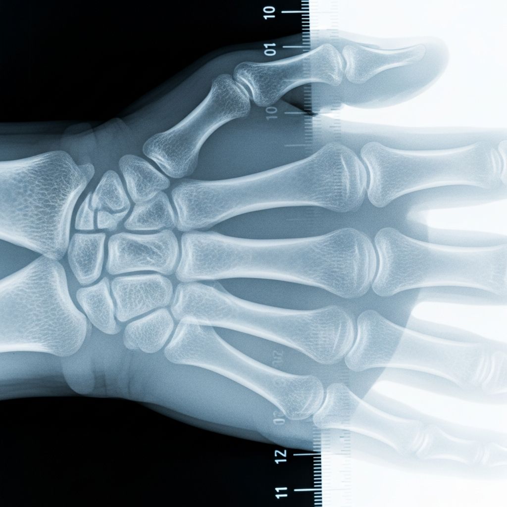
A bone age X-ray is a simple imaging test that gives doctors a snapshot of how mature your child’s bones are compared with most children the same age. It is commonly used when there are questions about growth, height, or the timing of puberty.
What Is Bone Age?
Bone age is an estimate of how far a child’s skeleton has developed, expressed in years, compared with typical patterns of bone growth for that age. In other words, it is a way to measure biological maturity, not just how many birthdays your child has had.
- Chronological age: the child’s age based on date of birth.
- Bone age (skeletal age): the age that best matches the appearance of the child’s bones on X-ray.
Bone age can be:
- Delayed – bones look younger than the child’s chronological age.
- Advanced – bones look older than expected for that age.
- Average – bones match what is typical for most children the same age.
How Does a Bone Age X-Ray Work?
To estimate bone age, doctors almost always use a single X-ray of the left hand and wrist. This area contains many small bones and growing zones that clearly show how mature the skeleton is.
Basic steps of the study
- Your child sits or stands beside the X-ray table.
- The left hand is placed flat on the plate, fingers comfortably spread.
- Your child stays still for 2–3 seconds while the image is taken.
- A pediatric radiologist or trained doctor reviews the X-ray and compares it to standard reference images.
The test uses a very small amount of ionizing radiation—similar to, or less than, many routine dental or limb X-rays—and is considered safe when medically indicated.
Comparing to reference atlases
To assign a bone age, the doctor compares your child’s X-ray to published collections of hand and wrist images from many children of known ages, such as the well-known Greulich and Pyle atlas.
| What the doctor looks at | What it means |
|---|---|
| Size and shape of the small bones in the hand | Shows how far the bones have lengthened and reshaped with age. |
| Appearance of growth plates (dark lines near the ends of bones) | Indicates how much growing potential remains; plates gradually thin and close over time. |
| Number of bones that have appeared (ossification centers) | Some bones are present only at certain stages, helping fine-tune the age estimate. |
What Are Growth Plates and Why Do They Matter?
Children’s long bones grow from special regions near their ends called growth plates (also known as epiphyseal plates). These are areas of softer cartilage that slowly turn into solid bone over many years.
- On X-ray, growth plates appear as darker lines between the main part of the bone and the ends.
- As a child matures, these plates become thinner.
- When growth is nearly complete, the plates “close,” blending into the rest of the bone.
Because growth plates follow a predictable timetable, their appearance is one of the key clues doctors use to estimate bone age and remaining growth potential.
Why Might a Doctor Order a Bone Age X-Ray?
Pediatricians and pediatric endocrinologists use bone age studies to answer questions about growth and development.
Common reasons to request the test
- Short stature – a child is much shorter than classmates or has dropped on the growth chart.
- Tall stature – a child is significantly taller than peers and growing faster than expected.
- Concerns about puberty timing – signs of puberty happening very early or much later than usual.
- Monitoring known hormone or genetic conditions – such as growth hormone deficiency, hypothyroidism, adrenal disorders, or Turner syndrome.
- Planning orthopedic or dental treatment – to time surgery, bracing, or jaw correction when growth will make treatment most effective.
How bone age helps in medical decisions
When interpreted alongside height, weight, growth records, family heights, and lab tests, bone age can help doctors:
- Estimate how much time a child has left to grow.
- Predict an approximate adult height range.
- Judge whether early or delayed puberty is likely to be temporary or linked to an underlying problem.
- Monitor how well treatments (for example, hormone therapy) are affecting growth.
What Happens During the Appointment?
A bone age X-ray visit is usually brief and straightforward. Children typically do not need sedation or special preparation.
Before the X-ray
- Your child can eat and drink normally.
- They may be asked to remove rings, watches, or bracelets from the left hand.
- Let the technologist know if your child has had any recent injuries to the hand or wrist.
During the X-ray
- The technologist positions your child at the machine and places the left hand flat on the X-ray surface.
- Your child is asked to hold still briefly while the picture is taken.
- The test is painless; your child will not feel the X-ray beam.
- If the first image is blurry from motion, a second quick image may be taken.
After the test
- Your child can resume normal activities right away.
- A pediatric radiologist or experienced doctor interprets the X-ray and sends a report to the ordering clinician.
- You will typically review the results at a follow-up visit or phone call with your child’s doctor.
Understanding the Results
The bone age report usually includes the estimated skeletal age and sometimes a brief description of how it compares with the child’s chronological age.
| Result pattern | Possible interpretation |
|---|---|
| Bone age ≈ chronological age | Growth appears to be following a typical timetable; many healthy children fall in this range. |
| Bone age < chronological age (delayed) | Skeleton looks younger than expected; may be seen in constitutional growth delay, some hormone disorders, chronic illnesses, or nutritional issues. |
| Bone age > chronological age (advanced) | Skeleton appears older than expected; may occur with early puberty, some adrenal or thyroid conditions, or certain genetic syndromes. |
It is important to remember:
- A difference between bone age and actual age does not automatically mean disease. Some healthy children naturally mature earlier or later than peers.
- The test gives an estimate, not an exact measurement; different readers may vary slightly in their assessments.
- Doctors always interpret bone age together with the child’s full medical history, physical exam, and other tests.
Conditions Where Bone Age Is Especially Useful
Bone age is a routine tool in pediatric endocrinology and radiology clinics around the world. It contributes to the evaluation and management of several conditions, including:
- Constitutional delay of growth and puberty
Children who are short in childhood but have delayed bone age and late puberty often catch up in height later than peers. - Growth hormone deficiency
Low growth hormone levels can slow bone maturation; bone age helps track response to therapy. - Hypothyroidism
Insufficient thyroid hormone in childhood can delay bone age and overall growth. - Precocious puberty
When puberty starts unusually early, bone age may be advanced, and growth plates can close sooner, shortening adult height without treatment. - Adrenal and sex steroid disorders
Excess or deficiency of hormones from the adrenal glands or gonads can affect skeletal maturation. - Genetic growth disorders (for example, Turner syndrome)
Bone age helps in long-term planning of growth and hormone therapies. - Orthopedic and orthodontic planning
Surgeons and orthodontists use bone age to time procedures so they align with growth spurts or the end of growth.
Is a Bone Age X-Ray Safe?
Bone age studies use low-dose X-rays. The amount of radiation is small and is generally considered safe when clearly needed for diagnosis or treatment decisions.
- X-rays have been used in children for many decades with well-established safety guidelines.
- Modern digital systems often use lower doses than older machines.
- Healthcare teams follow radiation-safety principles such as ALARA (As Low As Reasonably Achievable) to limit exposure.
If your child has had many imaging tests in the past, you can ask the doctor whether the bone age result will change management decisions. In many cases, the added information is valuable enough to justify this small exposure.
How Accurate Is Bone Age at Predicting Adult Height?
Doctors can combine bone age with your child’s current height, growth pattern, and family heights to estimate a likely adult height range. This is especially helpful when counseling families about early or delayed puberty and potential treatments.
However:
- Height predictions are estimates, not guarantees.
- Rapid changes in health, nutrition, hormones, or the timing of puberty can shift the final outcome.
- The younger the child, the wider the possible range of adult heights.
Your child’s doctor can explain how much confidence to place in the prediction for your specific situation.
Talking With Your Child About the Test
Many children feel more relaxed when they know what to expect. You can explain the bone age X-ray in simple terms:
- “The doctor wants to see how your bones are growing.”
- “They’ll take a quick picture of your hand, like a camera you can’t see.”
- “It doesn’t hurt, but you’ll need to keep your hand still for a few seconds.”
Reassure your child that the test is quick, you’ll be nearby, and it helps the doctor understand how to keep them healthy.
Frequently Asked Questions (FAQs)
How long does a bone age X-ray take?
The actual X-ray image is taken in just a few seconds. From check-in to leaving the imaging area, most families spend around 10–20 minutes, depending on the clinic’s schedule.
Does my child need any special preparation?
No fasting or special diet is needed. Your child may simply need to remove jewelry or metal items from the left hand and wrist.
Can a bone age test diagnose a condition by itself?
No. Bone age is one piece of the puzzle. Doctors interpret it alongside growth records, physical examination, family history, and laboratory tests to reach a diagnosis.
What if my child’s bone age is very different from their actual age?
A difference may point toward a growth pattern that deserves closer follow-up or further testing. However, some healthy children naturally mature early or late. Your pediatrician or pediatric endocrinologist can explain what it means in your child’s case.
Can bone age be repeated?
Yes. Doctors may repeat bone age studies over time to see how quickly the skeleton is maturing and to check how treatments are working. They balance the benefits of follow-up images against the small radiation exposure.
References
- Bone age — Wikipedia (summary of clinical methods, uses, and interpretation; accessed 2024-10-01). https://en.wikipedia.org/wiki/Bone_age
- X-Ray Exam: Bone Age Study — Nemours KidsHealth. 2023-05-10. https://kidshealth.org/en/parents/xray-bone-age.html
- Evaluation of Bone Age in Children: A Mini-Review — Frontiers in Pediatrics. 2021-02-09. https://www.frontiersin.org/articles/10.3389/fped.2021.580314
- Bone Age — StatPearls, NCBI Bookshelf. Updated 2022-07-11. https://www.ncbi.nlm.nih.gov/books/NBK537051/
- What Is Bone Age? How X-Rays Reveal Your Child’s Growth Timeline — Life Pediatric Endocrinology Blog. 2023-03-01. https://lifeendo.com/blog/bone-age-xray-growth-puberty