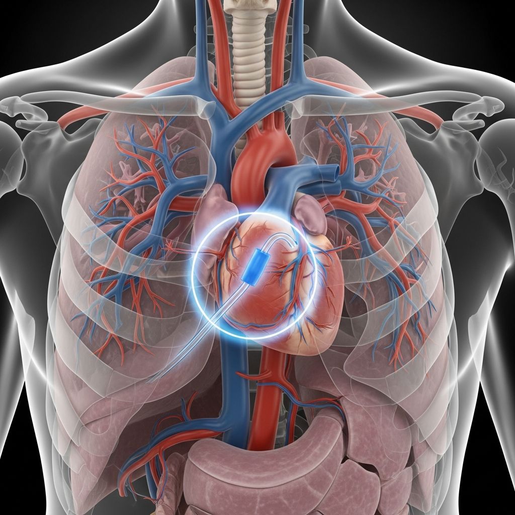
The Blalock-Taussig-Thomas (BTT) shunt, often simply called the BT shunt, is a palliative surgical intervention that creates an artificial pathway to boost blood flow to the lungs in infants with certain congenital heart defects. This procedure dramatically improves oxygenation, alleviating the life-threatening cyanosis—bluish skin tint from oxygen deprivation—that affects these vulnerable babies.
Pioneering Origins of a Medical Breakthrough
The story of the BT shunt begins in the 1940s amid desperate efforts to save ‘blue babies’ dying from inadequate pulmonary circulation. In 1944, at Johns Hopkins Hospital, surgeon Dr. Alfred Blalock, pediatric cardiologist Dr. Helen Taussig, and lab technician Vivien Thomas collaborated on the first successful operation. Thomas’s meticulous animal experiments refined the technique of connecting the subclavian artery to the pulmonary artery, directly inspiring the human procedure performed on November 29, 1944.
Originally named the Blalock-Taussig shunt, it was later updated to Blalock-Taussig-Thomas to honor Thomas’s pivotal role, which had been overlooked for decades due to racial biases of the era. This innovation marked the dawn of modern pediatric cardiac surgery, proving that complex heart anomalies in newborns could be surgically palliated.
When Is a BT Shunt Necessary?
BT shunts are indicated for cyanotic congenital heart diseases (CHDs) where pulmonary blood flow is severely restricted. Common conditions include:
- Tetralogy of Fallot: A quartet of defects causing right ventricular outflow obstruction and ventricular septal defect.
- Pulmonary Atresia: Complete blockage of the pulmonary valve, preventing blood from reaching the lungs.
- Hypoplastic Left Heart Syndrome (HLHS): Underdeveloped left heart structures reliant on ductal flow.
- Tricuspid Atresia: Absent or imperforate tricuspid valve blocking right atrial emptying.
- Severe Pulmonary Stenosis: Narrowing that drastically limits lung-directed blood.
These defects often manifest in the first days of life as the ductus arteriosus—a fetal vessel—closes, plunging oxygen saturation levels. Without intervention, infants face rapid deterioration, hypoxia, and death.
Evolution: From Classic to Modified BT Shunt
The classic BT shunt involved directly anastomosing (sewing end-to-end) the subclavian artery to the pulmonary artery via thoracotomy. This sacrificed arm blood supply and risked arm ischemia, plus pulmonary artery distortion as the child grew.
The modified Blalock-Taussig-Thomas (mBTT) shunt, introduced later, uses a synthetic polytetrafluoroethylene (PTFE) graft—typically 3.5-5mm diameter—interposed between the subclavian or innominate artery and the right pulmonary artery. This preserves native vessel integrity, allows adjustable sizing, and minimizes complications.
| Aspect | Classic BT Shunt | Modified BTT Shunt |
|---|---|---|
| Surgical Material | Direct artery-to-artery anastomosis | PTFE synthetic tube |
| Approach | Lateral thoracotomy | Median sternotomy |
| Arm Blood Supply | Often compromised | Preserved |
| Bypass Use | Usually off-pump | Flexible (on/off-pump) |
| Long-term Growth | Distortion risk | Better accommodation |
This table highlights key advancements making mBTT safer and more versatile.
Detailed Surgical Procedure
Performed in neonates (often first 1-2 weeks), the mBTT shunt surgery lasts 2-4 hours under general anesthesia. Surgeons favor median sternotomy for superior exposure and emergency access.
Steps include:
- Incision and Exposure: Midline chest incision; partial thymectomy; pericardiotomy to visualize great vessels.
- Vessel Prep: Isolate subclavian/innominate artery and right pulmonary artery (RPA); apply vascular clamps.
- Graft Anastomosis: Bevel PTFE ends; sew to artery (end-to-side) and RPA with fine polypropylene sutures for hemostasis.
- Flow Assessment: Release clamps; confirm shunt patency via Doppler or direct flow.
- Closure: Chest tubes, pacing wires, sternal wiring; heparin reversal optional.
Many centers operate off-pump, but bypass supports complex cases.
Immediate Postoperative Care
Infants recover in cardiac ICU with mechanical ventilation initially. Monitoring focuses on:
- Stable hemodynamics and oxygen saturation (>75-85%).
- Shunt patency (continuous pulse oximetry, echo).
- Preventing thrombosis (heparin infusion, lifelong low-dose aspirin).
Most wean from ventilator in 1-3 days; ICU stay 5-10 days; hospital discharge 1-3 weeks. Complications (10-20%) include shunt occlusion (2-5%), over-shunting (pulmonary hypertension), bleeding, infection, or phrenic nerve injury (diaphragm paralysis).
Long-Term Role in Staged Repair
The BT shunt is temporary (3-12 months), buying time for pulmonary artery growth and heart conditioning before corrective surgeries like Glenn or Fontan in single-ventricle palliation, or full TOF repair.
Takedown occurs during definitive operation: ligate/clamp shunt, resect graft, repair pulmonary artery distortion (‘tenting’). Survival post-shunt exceeds 90% in expert centers, with reduced cyanosis and improved growth.
Risks, Complications, and Outcomes
While transformative, risks persist:
- Shunt Thrombosis: 5-10%; mitigated by antiplatelets, optimal sizing.
- Overcirculation: Heart strain from excess flow.
- Infection/Arrhythmia: Rare but serious.
Modern outcomes: >95% procedural survival; bridges 80-90% to next-stage surgery. Developing nations rely heavily on it due to resource limits.
Life After the Shunt: Family Guidance
Post-discharge, parents administer aspirin, monitor color/breathing, avoid infections, and attend frequent cardiology visits. Activity ramps up gradually; feeds improve with better oxygenation. Emotional support is vital—connect with CHD families.
Frequently Asked Questions (FAQs)
How soon after birth is BT shunt surgery performed?
Typically within the first 1-2 weeks, as ductus closure worsens cyanosis.
Does the shunt fix the heart defect permanently?
No, it’s palliative; full repair follows later.
What signs indicate shunt problems?
Increasing blueness, lethargy, poor feeding, low oxygen sats—seek emergency care.
Will my child need medications lifelong?
Aspirin yes; others depend on underlying defect and repairs.
Can BT shunt patients lead normal lives?
With staged surgeries, many thrive into adulthood with good quality of life.
Future Directions in Shunt Therapy
Though declining in high-resource settings due to primary neonatal repairs, mBTT remains cornerstone for univentricular hearts and resource-poor areas. Innovations like absorbable grafts or hybrid stenting may further evolve options.
References
- Modified Blalock-Taussig-Thomas Shunt – StatPearls — NCBI Bookshelf/StatPearls Publishing. 2023-07-17. https://www.ncbi.nlm.nih.gov/books/NBK597363/
- Blalock-Taussig shunt | Stanford Health Care — Stanford Health Care. 2023. https://stanfordhealthcare.org/medical-conditions/blood-heart-circulation/single-ventricle/treatments/blalock-taussig-shunt.html
- Blalock-Taussig (BT) Shunt – Norton Children’s — Norton Children’s Heart Institute. 2023. https://nortonchildrens.com/services/cardiology/conditions/treatments/surgical/blalock-taussig-bt-shunt/
- Blalock-Taussig-Thomas (BTT) Shunt – Cleveland Clinic — Cleveland Clinic. 2023-11-01. https://my.clevelandclinic.org/health/procedures/btt-shunt
- Pediatric Blalock-Taussig shunt – Children’s Health — Children’s Health. 2023. https://www.childrens.com/specialties-services/treatments/blalock-taussig-shunt
- New name for lifesaving procedure recognizes Black pioneer’s role — American Medical Association. 2021-12-14. https://www.ama-assn.org/public-health/health-equity/new-name-lifesaving-procedure-recognizes-black-pioneer-s-role