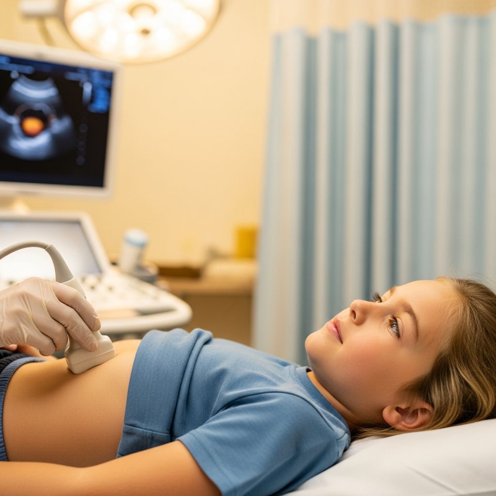
When a doctor needs a closer look at a child’s urinary system, a bladder ultrasound is often the first test they choose. It is noninvasive, does not use radiation, and is well suited for babies, toddlers, and older children alike.
This guide explains in simple language why a bladder ultrasound might be ordered, how to prepare your child, what happens during the exam, and what the results can tell your child’s care team.
What Is a Bladder Ultrasound?
A bladder ultrasound (often done together with a kidney or “renal” ultrasound) is an imaging test that uses sound waves to create pictures of the bladder and nearby urinary structures.
- No radiation: Unlike X-rays or CT scans, ultrasound uses sound waves, not ionizing radiation.
- Real-time images: The machine shows moving pictures of the bladder filling and emptying.
- Safe for children: Ultrasound has been used safely in pediatric care for decades and is considered ideal for urinary tract imaging in kids.
The test is sometimes called a renal–bladder ultrasound because the kidneys and bladder are often checked during the same visit.
Why Might a Child Need a Bladder Ultrasound?
Doctors order bladder ultrasounds for many different reasons. Common situations include:
- Frequent urinary tract infections (UTIs)
- Pain or burning with urination
- Blood in the urine (hematuria)
- Trouble starting to pee or fully emptying the bladder
- Daytime accidents or bedwetting, especially when new or severe
- Abdominal or side (flank) pain the doctor thinks may be related to the urinary tract
- Abnormal prenatal ultrasound suggesting kidney or bladder differences
- Follow-up after surgery or treatment for urinary problems
| Possible Issue | How Ultrasound Helps |
|---|---|
| Structural differences (congenital anomalies) | Shows the size, shape, and position of the kidneys, ureters, and bladder. |
| Bladder wall thickening | Measures the bladder wall and checks for inflammation or obstruction. |
| Incomplete bladder emptying | Compares bladder before and after urination (pre- and post-void images). |
| Hydronephrosis or urinary tract dilation | Looks for backup of urine into the kidneys or ureters. |
| Masses, stones, or debris | Shows stones, tumors, or thick material in the bladder or kidneys. |
How Ultrasound Works (In Kid-Friendly Terms)
Ultrasound machines send out high-frequency sound waves from a small handheld device called a transducer. These sound waves bounce off tissues and organs, and a computer turns the echoes into images on a screen.
- Your child cannot feel the sound waves.
- The only sensation is the cool or slightly warm gel and gentle pressure from the probe.
- No needles or injections are needed for a standard bladder ultrasound.
Preparing Your Child for a Bladder Ultrasound
Preparation is usually simple, but it can make a big difference in image quality and how smoothly the visit goes.
Bladder Filling Instructions
For many kidney and bladder ultrasounds, the imaging team prefers a full bladder at the beginning of the exam.
- Older children may be asked to drink water 30–60 minutes before the test and avoid urinating until told.
- Younger children may be given fluids in the waiting area; the technologist will time the exam as the bladder fills.
- If the bladder is not full enough, the sonographer may pause and ask your child to drink more before continuing.
Follow any instructions your hospital or imaging center provides, as preparation can vary slightly based on age and the exact study needed.
Clothing and Comfort
- Dress your child in two-piece outfits so the abdomen can be easily exposed.
- Leave jewelry at home if it might get in the way of the scan.
- Bring a favorite toy, blanket, book, or device to help your child relax while lying still.
Emotional Preparation
Children often feel calmer when they know what to expect. Consider:
- Explaining that the test uses a “camera” that looks inside the body using sound.
- Describing the gel as “warm jelly” or “slippery lotion.”
- Reassuring them that it should not hurt, though they may feel gentle pressing.
- Practicing taking deep breaths or staying still for short periods at home.
Step-by-Step: What Happens During the Exam
Most pediatric bladder ultrasounds are completed in about 15–30 minutes, depending on your child’s age, cooperation, and how quickly the bladder fills.
1. Check-in and Brief Interview
- You and your child check in at the imaging department.
- A technologist (sonographer) may ask about your child’s symptoms and reason for the exam.
- They will confirm any special instructions (such as a full bladder or recent void).
2. Positioning on the Exam Table
- Your child lies on a padded table, usually on their back.
- Parents are often encouraged to stay in the room for reassurance, unless pregnancy or other policies limit this.
3. Applying Gel and Placing the Probe
- The sonographer puts clear, water-based gel on the child’s lower abdomen (and sometimes the sides or back).
- The gel helps sound waves travel better and prevents air pockets.
- The technologist slides the probe over the area, watching the screen in real time.
4. Images with a Full Bladder
Images are usually taken while the bladder is reasonably full to see its shape and wall clearly and to check the ureters where they enter the bladder.
- The technologist takes pictures in different directions (longways and crossways).
- Your child may be asked to take a few deep breaths or hold still briefly, especially if the kidneys are also being scanned.
5. Going to the Bathroom (If Needed)
For many children who can use the toilet, the team will want to compare the bladder before and after urination (pre- and post-void images).
- The technologist will tell your child when they may go pee.
- Afterward, your child returns to the table for more quick pictures of the “emptied” bladder.
6. After the Scan
- The sonographer checks that they have all needed images before wiping away the gel.
- There are no activity or diet restrictions from a standard bladder ultrasound; your child can return to normal right away.
- A radiologist or pediatric imaging doctor reviews the images and sends a report to your child’s referring provider.
Is a Bladder Ultrasound Safe?
Diagnostic ultrasound is considered very safe and is widely used in children, including newborns.
- No ionizing radiation exposure, unlike X-ray or CT scans.
- Most examinations do not require sedation or anesthesia.
- There are no known long-term side effects from routine medical ultrasound at approved settings.
Some children may find the pressure from the probe mildly uncomfortable, especially if the bladder is very full, but this can usually be adjusted by the technologist.
Understanding Possible Findings
Your child’s doctor will interpret the ultrasound results in the context of symptoms, physical exam, and any laboratory tests.
Examples of What the Report May Describe
- Bladder size and shape: Whether the bladder appears normal for age and how full it was at the time of the scan.
- Bladder wall: Thickness and smoothness of the wall; significant thickening can suggest obstruction or chronic irritation.
- Residual urine volume: How much urine remains after your child voids; small amounts (around 20 mL or less) are often considered within normal limits, depending on age.
- Kidneys and ureters: Whether there are any signs of swelling (hydronephrosis), stones, or other changes.
- Other visible structures: Sometimes nearby organs or tissues are also seen and mentioned.
Abnormal results do not automatically mean serious disease. In many cases, the ultrasound helps rule out major problems and guides the next steps, which might include observation, repeat imaging, or additional tests.
Special Types of Ultrasound Involving the Bladder
In some situations, doctors may recommend more specialized ultrasound procedures that focus on how urine flows.
Voiding Studies Using Ultrasound
One such test is contrast-enhanced voiding urosonography (ceVUS), which evaluates for urine flowing backward from the bladder to the kidneys (vesicoureteral reflux).
- A small catheter is placed into the urethra and contrast agent diluted in saline is run into the bladder.
- Ultrasound images are taken while the bladder fills and while the child urinates.
- The contrast makes it easier to see whether urine is traveling in the wrong direction toward the kidneys.
Unlike traditional X-ray voiding cystourethrograms, ceVUS uses ultrasound instead of fluoroscopy, avoiding radiation exposure while still providing detailed views of reflux.
Helping Your Child Cope with the Experience
Parents play an important role in keeping children calm and cooperative during imaging.
- Stay close: Hold your child’s hand, talk softly, or help distract them with stories or music.
- Use honest reassurance: Acknowledge any worries and emphasize that the test is to help the doctor keep them healthy.
- Ask about distractions: Many pediatric imaging departments offer videos, toys, or games during the scan.
- Plan a reward: A small treat or special activity afterward can give your child something positive to look forward to.
Frequently Asked Questions (FAQs)
Will my child feel pain during a bladder ultrasound?A: The test itself is not painful. Your child may feel cool or warm gel and light pressure from the probe. If the bladder is very full, they may feel some temporary discomfort, but this usually improves once they are allowed to urinate.
Q: How long does the exam usually take?
For most children, the scan takes about 15–30 minutes. It can be longer if the bladder is not full at the start or if the child has difficulty lying still.
Can my child eat and drink normally before the test?
In many cases, yes. Some centers will give specific instructions, such as drinking water beforehand to fill the bladder, but most bladder ultrasounds do not require changes in diet.
When will we get the results?
A radiologist or pediatric imaging specialist reviews the images and sends a written report to the doctor who ordered the test. Timing varies, but many families receive results within a few days, sometimes sooner depending on the urgency and the facility’s process.
Is this test enough to diagnose all urinary problems?
Ultrasound provides valuable information about structure and bladder function, but it may not answer every question. Depending on the findings and your child’s symptoms, the doctor may recommend additional tests, lab work, or follow-up imaging.
Does ultrasound expose my child to radiation?
No. Ultrasound uses sound waves, not ionizing radiation, which is one reason it is strongly preferred for initial assessment of the urinary tract in children.
References
- Pediatric Contrast-Enhanced Voiding Urosonography (ceVUS) — Radiological Society of North America / RadiologyInfo.org. 2022-10-18. https://www.radiologyinfo.org/en/info/cevus
- Renal Bladder Ultrasound — UCSF Department of Urology. 2013-01-23. https://urology.ucsf.edu/sites/default/files/uploaded-files/basic-page/renal_bladder_ultrasound.pdf
- Children’s (Paediatric) Renal Ultrasound — InsideRadiology / Royal Australian and New Zealand College of Radiologists. 2016-02-01. https://www.insideradiology.com.au/paediatric-renal-ultrasound/
- A practical guide to urinary tract ultrasound in a child — Riccabona M et al., Insights into Imaging (Springer). 2016-02-15. https://pmc.ncbi.nlm.nih.gov/articles/PMC4760558/
- Urosonography (ceVUS) — Children’s Hospital Colorado. 2023-05-10. https://www.childrenscolorado.org/doctors-and-departments/departments/imaging-services/cevus/
- Ultrasound of the kidney, bladder, or pelvis for children and adults — University of Iowa Hospitals & Clinics. 2022-04-20. https://uihc.org/educational-resources/ultrasound-kidney-bladder-or-pelvis-children-and-adults
- Pediatric Ultrasound — Children’s Hospital of Philadelphia. 2021-09-01. https://www.chop.edu/treatments/ultrasound