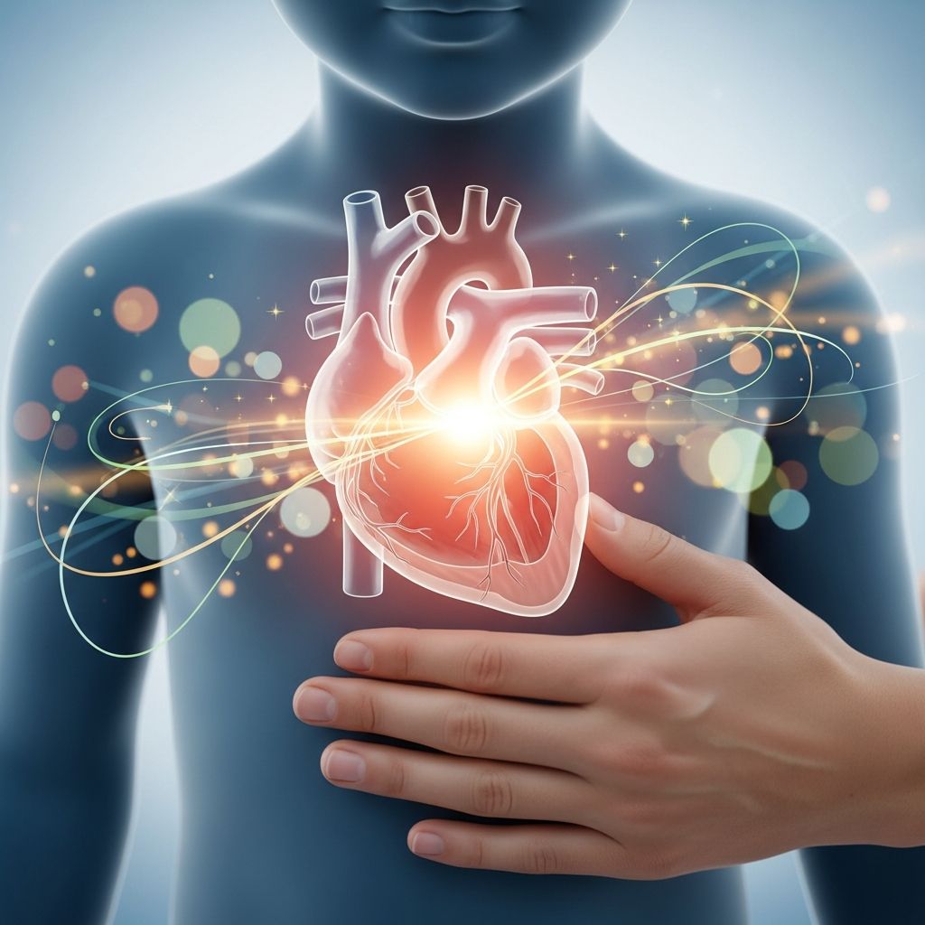
Atrioventricular canal defect, commonly called AV canal defect, represents a significant congenital heart anomaly where the central heart partition fails to develop correctly during fetal growth. This condition disrupts normal blood circulation by permitting mixing of oxygenated and deoxygenated blood, while also involving malformed valves between the heart’s upper and lower sections.
Understanding the Anatomy of AV Canal Defect
The human heart features four chambers: two atria on top and two ventricles below, separated by septa to ensure efficient blood flow. In a typical setup, oxygen-depleted blood enters the right atrium, moves to the right ventricle, and heads to the lungs for oxygenation. Oxygen-rich blood then returns to the left atrium, passes through the left ventricle, and circulates to the body. Valves—the tricuspid on the right and mitral on the left—prevent backward flow.
In AV canal defect, the endocardial cushions, tissues that form the central septum and valves during embryonic stages, do not fuse properly. This results in a sizable gap bridging the atrial and ventricular septa, often accompanied by a single oversized valve instead of distinct ones. Consequently, blood shunts abnormally, overfilling the lungs and under-oxygenating systemic circulation.
Types of Atrioventricular Canal Defects
AV canal defects vary in severity, classified primarily as partial or complete, with transitional forms in between.
- Partial AV Canal Defect: Features a hole solely in the atrial septum (primum atrial septal defect) alongside deformed mitral and tricuspid valves that leak. Ventricular separation remains intact, leading to milder mixing.
- Complete AV Canal Defect: Involves holes in both atrial and ventricular septa, forming a single large central opening. A common atrioventricular valve bridges all chambers, causing substantial blood mixing and pulmonary overcirculation.
- Transitional Forms: Small ventricular septal components with larger atrial defects and valve issues, behaving between partial and complete types.
These distinctions influence symptom onset and intervention timing, with complete variants demanding earlier action.
Prevalence and Associated Risk Factors
Accounting for 3-5% of congenital heart diseases, AV canal defects affect about 1 in 2,000-3,000 live births. Notably, around 15-20% of Down syndrome (trisomy 21) infants exhibit this defect, far exceeding general rates due to chromosomal impacts on heart development. Other genetic syndromes and maternal factors like diabetes or rubella exposure elevate risk, though most cases arise sporadically.
| Risk Factor | Prevalence Impact | Source |
|---|---|---|
| Down Syndrome | 15-20% of cases | |
| General Population | 0.03-0.05% of births | |
| Maternal Diabetes | Increased odds |
Recognizing Symptoms in Infants and Children
Manifestations depend on defect magnitude. Partial types may remain asymptomatic initially, detected incidentally. Complete defects provoke early signs from excessive pulmonary blood flow.
- Poor feeding and slow weight gain due to fatigue during meals.
- Rapid breathing or breathlessness, especially when upright.
- Frequent lung infections like pneumonia.
- Bluish skin tint (cyanosis) around lips or extremities in advanced mixing.
- Excessive sweating, particularly on the head during activity.
Untreated, these progress to heart failure: enlarged heart, fluid retention, and liver congestion. Pulmonary hypertension may develop from sustained high lung pressures, risking irreversible vascular damage (Eisenmenger syndrome).
Diagnostic Approaches for AV Canal Defect
Prenatal ultrasound often identifies the defect via abnormal four-chamber heart views. Postnatally, pediatricians note murmurs—turbulent flow sounds—prompting further tests.
- Echocardiogram: Gold standard, visualizing septal gaps, valve function, and shunting direction.
- Chest X-ray: Reveals cardiomegaly and lung congestion.
- ECG: Shows atrial enlargement or conduction issues.
- Cardiac MRI/Catheterization: For complex cases assessing pulmonary pressures.
Early diagnosis facilitates timely surgery, averting complications like endocarditis from turbulent flows on irregular valves.
Surgical Interventions: Repairing the Heart
Definitive treatment entails open-heart surgery, ideally within the first 3-6 months for complete defects to forestall pulmonary issues. Procedures occur under cardiopulmonary bypass.
- Septal patching with synthetic or pericardial material to seal ASD/VSD.
- Valve reconstruction: Dividing the common valve into tricuspid/mitral components, often with annuloplasty.
- Addressing associated anomalies like left ventricular outflow issues.
Success rates surpass 95% in premier centers, though reoperations occur in 10-20% for valve leaks or residual shunts later in life.
Post-Surgical Care and Recovery
Immediate post-op includes ICU monitoring for arrhythmias, bleeding, or low output. Hospital stays span 7-14 days, with activity restrictions for 4-6 weeks. Medications like diuretics, ACE inhibitors, or antiarrhythmics aid recovery.
Long-term, patients require endocarditis prophylaxis, routine echoes, and lifestyle adjustments: low-sodium diet, exercise moderation, and Down syndrome screenings if applicable. Pregnancy counseling is vital for females due to heightened risks.
Potential Complications and Long-Term Outlook
While most lead active lives post-repair, vigilance is key. Valve regurgitation, conduit narrowing, or arrhythmias may necessitate interventions. Adults face higher pulmonary hypertension risks than simpler defects. Regular cardiology follow-up ensures early detection.
Prognosis excels with modern care: 90%+ survival to adulthood, many athletically capable sans restrictions.
Special Considerations for Children with Down Syndrome
Given elevated incidence, AV canal vigilance is routine in Down syndrome newborns. These children tolerate surgery well but contend with pulmonary vulnerabilities and other anomalies. Multidisciplinary teams—including genetics, pulmonology—optimize outcomes.
Emotional Support for Families
A diagnosis overwhelms; connect with groups like Mended Little Hearts or CHD Ireland for peer support. Educate on condition empowers advocacy during treatments. Financial planning aids surgical costs.
Frequently Asked Questions About AV Canal Defect
Can AV canal defect be detected before birth?
Yes, fetal echocardiography around 18-22 weeks often spots it, enabling delivery planning at specialized centers.
Is surgery always necessary?
Yes for complete forms; partial may watch if mild, but most require repair to avert failure.
What is the recovery like after heart surgery?
Initial hospital time, gradual activity return; full recovery in months with follow-ups.
Do children outgrow AV canal defect?
No, it demands surgical correction; growth doesn’t resolve structural flaws.
Are there activity limits post-repair?
Contact sports avoided initially; most resume normalcy with clearance.
Preventive Measures and Ongoing Monitoring
While unpreventable, folic acid in pregnancy cuts CHD risks broadly. Post-repair, annual assessments track valve function, ventricular size, and pressures. Vaccinations against respiratory illnesses protect healing lungs.
AV canal defect, though formidable, yields to expertise. Informed parents partnering with cardiologists secure brighter futures for their children.
References
- AV Canal: Atrioventricular Canal Defect Surgery — Rady Children’s Hospital San Diego. 2023. https://www.rchsd.org/programs-services/heart-institute/services-conditions-treated/av-canal-defect/
- Complete Atrioventricular Canal Defect (CAVC) — American Heart Association. 2024-01-15. https://www.heart.org/en/health-topics/congenital-heart-defects/about-congenital-heart-defects/complete-atrioventricular-canal-defect-cavc
- Atrioventricular Canal Defect — Nemours KidsHealth. 2025. https://kidshealth.org/en/parents/av-canal-defect.html
- Atrioventricular Canal Defect – Symptoms & Causes — Mayo Clinic. 2024-06-10. https://www.mayoclinic.org/diseases-conditions/atrioventricular-canal-defect/symptoms-causes/syc-20361492
- Atrioventricular Canal Defect — Children’s Hospital of Orange County (CHOC). 2024. https://choc.org/conditions/cardiology/atrioventricular-canal-defect/
- Atrioventricular Canal (Septal) Defect — Cleveland Clinic. 2025-01-03. https://my.clevelandclinic.org/health/diseases/22128-atrioventricular-canal-defect