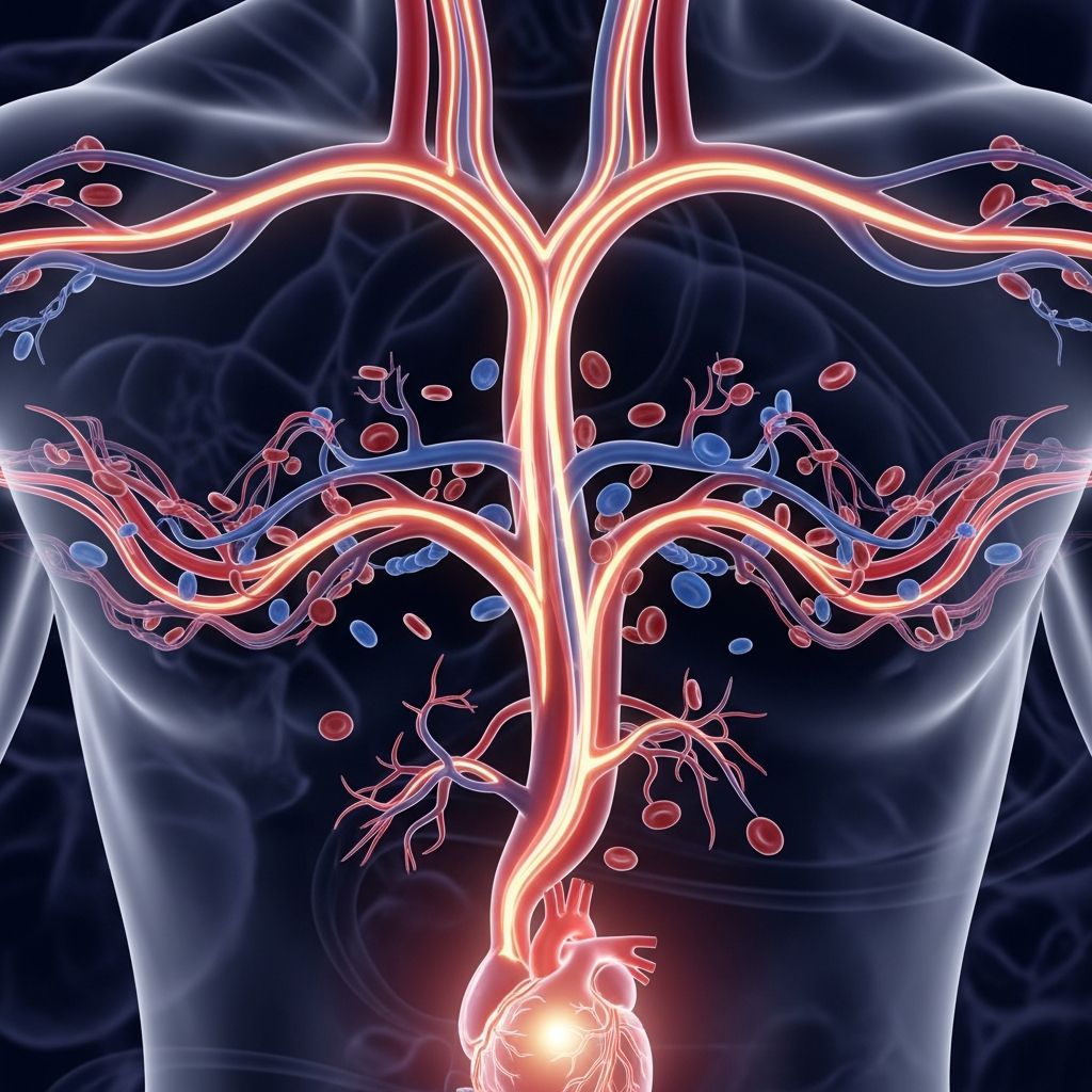
What Is the Aorta?
The aorta stands as the body’s largest and most important artery, serving as the primary conduit through which the heart pumps oxygen-rich blood throughout the entire body. This remarkable vessel originates directly from the left ventricle of the heart and extends downward through the chest and abdomen, ultimately splitting into smaller arteries that distribute nutrients and oxygen to virtually every cell in the body. Without the aorta functioning properly, the body cannot deliver the essential oxygen needed for organs, tissues, and cells to survive even for a few minutes.
The aorta’s impressive dimensions reflect its critical responsibility. This vessel measures more than one foot in length and can be over one inch in diameter at its widest point, making it substantially larger than any other blood vessel in the circulatory system. The vessel possesses exceptionally thick walls designed to withstand the considerable pressure generated by each heartbeat, ensuring it can safely transport blood without rupturing or collapsing.
Structural Composition and Layers
The aorta’s strength and durability stem from its complex, multi-layered construction. The arterial wall consists of three distinct tissue layers, each serving specific functions to maintain the vessel’s integrity and performance.
- The Inner Layer (Intima): This innermost lining comes into direct contact with flowing blood and maintains a smooth surface that promotes efficient blood movement without unnecessary friction.
- The Middle Layer (Media): Composed primarily of muscle tissue and elastic fibers, this thick muscular layer enables the aorta to expand when receiving blood from the heart and contract as the vessel propels blood forward throughout the circulatory cycle.
- The Outer Layer (Adventitia): This protective outer covering provides structural support and connects the aorta to surrounding tissues, anchoring it securely within the chest and abdominal cavities.
Beyond these primary layers, the aorta receives nourishment through an extensive network of tiny blood vessels called the vasa vasorum. These microscopic vessels supply oxygen and nutrients to the aortic wall itself, much like the coronary arteries feed the heart muscle. This intricate blood supply becomes increasingly important in maintaining aortic health, particularly as the vessel ages.
Anatomical Divisions and Pathways
The aorta does not travel as a single, uniform tube through the body. Instead, it follows a specific pathway with distinct sections, each named according to its location and the organs it supplies.
The Ascending Aorta
The ascending aorta begins at the aortic valve, the gateway through which blood exits the left ventricle. This initial segment extends upward approximately five centimeters toward the shoulders and neck. The ascending aorta has critical importance because the coronary arteries branch from this region, delivering oxygen directly to the heart muscle itself. These branches ensure the heart continues beating with sufficient oxygen to support its own function.
The Aortic Arch
As the ascending aorta reaches the top of the chest, it curves in a distinctive arc known as the aortic arch. This curved section acts as a major distribution point, with three significant branches emerging to supply blood to the head, neck, arms, and brain. The arch region contains specialized nerve endings called baroreceptors and chemoreceptors that monitor blood pressure and oxygen levels, sending this information back to the brain to help regulate cardiovascular function.
The Thoracic Descending Aorta
After completing its arch, the aorta descends through the chest cavity as the thoracic descending aorta. This section supplies blood to the chest structures, including the esophagus, lungs, and surrounding tissues. Numerous smaller branches extend from this region to serve the vital structures within the thoracic cavity.
The Abdominal Aorta
The aorta continues its downward journey as it passes through the diaphragm and enters the abdominal cavity, where it becomes the abdominal aorta. This final major section represents the largest and longest portion of the aorta and supplies critical organs including the stomach, intestines, kidneys, liver, and spleen. The abdominal aorta eventually terminates by dividing into two smaller arteries called the common iliac arteries, which then distribute blood to the pelvis and lower limbs.
Blood Distribution and Organ Supply
Understanding which organs and body regions receive blood from specific aortic segments provides insight into why aortic disease can be so dangerous. If blood flow through any section becomes compromised, the organs that depend on that segment face immediate oxygen deprivation.
| Aortic Section | Primary Organs and Tissues Supplied |
|---|---|
| Ascending Aorta | Heart muscle via coronary arteries |
| Aortic Arch | Brain, head, neck, arms, and shoulders |
| Thoracic Descending Aorta | Lungs, esophagus, chest wall, and mediastinal structures |
| Abdominal Aorta | Stomach, intestines, kidneys, liver, spleen, and surrounding abdominal organs |
| Common Iliac Arteries (Terminal Branches) | Pelvis, buttocks, and lower limbs |
The aorta’s extensive branching pattern ensures that every region of the body receives the oxygenated blood necessary for survival. This comprehensive distribution network distinguishes the aorta from other blood vessels and explains why cardiovascular surgeons consider it the foundation of the entire circulatory system.
The Aorta’s Critical Functional Role
The aorta’s primary responsibility involves receiving blood freshly pumped from the left ventricle and distributing it throughout the body via its numerous branches and subdivisions. Each heartbeat generates a surge of pressure that pushes oxygen-rich blood into the aorta, which then propels this vital fluid forward through increasingly smaller branching arteries until it reaches individual cells.
Beyond oxygen delivery, the aorta facilitates the transport of essential nutrients, hormones, and other vital substances throughout the body. The endothelial lining (inner layer) of the aorta maintains an incredibly smooth surface that minimizes resistance to blood flow, allowing the heart to pump more efficiently. This efficient transport system has evolved over millions of years to support human life with remarkable reliability and durability.
The aorta’s muscular middle layer provides additional functionality by expanding elastically during systole (when the heart contracts) and recoiling during diastole (when the heart relaxes). This elastic recoil helps propel blood forward even between heartbeats, smoothing out the pulsatile nature of cardiac output and maintaining steady blood pressure throughout the circulatory system.
Why the Aorta Requires Special Protection
Because the aorta represents the single most critical vessel in the human body, it has evolved multiple protective mechanisms and specialized features that other arteries lack. The thick muscular walls prevent rupture despite operating under considerable pressure continuously throughout life. The surrounding pericardial sheath and proximity to the rib cage provide physical protection from external trauma.
Additionally, the network of baroreceptors and chemoreceptors embedded within the aortic arch provides real-time feedback to the brain about cardiovascular status, allowing the nervous system to make continuous adjustments to heart rate and blood vessel diameter to maintain optimal blood pressure and oxygenation. This sophisticated monitoring system ensures the aorta can adapt to changing physiological demands, whether during rest, physical exercise, or stress.
Common Aortic Conditions and Health Concerns
Despite its impressive design, the aorta remains vulnerable to disease processes that can develop gradually over time. Atherosclerosis, characterized by the buildup of fatty deposits on arterial walls, commonly affects the descending aorta and can reduce blood flow to critical organs. High blood pressure, the most frequent cause of aortic disease in the abdominal segment, can weaken the arterial wall over decades. Other conditions such as aortic aneurysms (bulging sections of the weakened wall) or aortic dissection (tearing of the inner lining) represent life-threatening emergencies requiring immediate medical intervention.
The infrarenal abdominal aorta (the section below the kidney arteries) carries particular risk for aneurysm formation because this region receives reduced blood supply from the vasa vasorum compared to the thoracic aorta. This reduced nutritional supply to the aortic walls can increase vulnerability to inflammation and degeneration over time.
Maintaining Aortic Health Throughout Life
Protecting aortic function involves adopting cardiovascular health strategies that apply across the lifespan. Maintaining healthy blood pressure through regular physical activity, appropriate sodium intake, and stress management reduces wear on the aortic walls. Managing cholesterol levels helps prevent atherosclerotic buildup that can narrow the aorta and reduce blood flow. Avoiding tobacco smoke, limiting alcohol consumption, and maintaining a healthy body weight all contribute to long-term aortic health.
Regular medical check-ups, particularly for individuals with family histories of aortic disease or those with risk factors like diabetes or chronic hypertension, enable early detection of aortic problems before they progress to critical stages. Modern imaging technologies including ultrasound, CT scans, and MRI allow physicians to monitor aortic diameter and identify concerning changes.
Frequently Asked Questions About the Aorta
How long does it take for blood to travel through the entire aorta?
Blood typically circulates through the aorta and returns to the heart within approximately one minute during rest. During physical exertion, circulation accelerates significantly as heart rate increases.
Can the aorta heal itself if damaged?
While the aorta possesses some capacity for repair through vascular remodeling, significant damage such as dissection or large aneurysms typically requires medical intervention. Minor atherosclerotic changes may stabilize with aggressive risk factor management, but advanced disease usually requires surgical or endovascular treatment.
What symptoms might indicate aortic problems?
Aortic dissection presents with sudden, severe chest or back pain. Aortic aneurysms often develop silently without symptoms until they rupture. Some people experience abdominal or back pain, pulsatile sensations, or shortness of breath. Any sudden severe chest or back pain warrants emergency medical evaluation.
How do doctors diagnose aortic disease?
Physicians use imaging studies including echocardiography, CT angiography, MRI, and transesophageal echocardiography to visualize the aorta and identify abnormalities. Physical examination may reveal findings suggestive of aortic problems, prompting further imaging investigation.
Is exercise safe if someone has an aortic condition?
Exercise recommendations depend on the specific aortic condition and its severity. Some patients benefit from supervised, moderate-intensity exercise, while those with large aneurysms or dissections may require activity restrictions. Cardiologists should provide individualized guidance based on diagnostic findings.
References
- Aorta: Anatomy and Function — Cleveland Clinic. 2024. https://my.clevelandclinic.org/health/body/17058-aorta-anatomy
- Aorta Health: Risks and Symptoms of Aneurysm — American Heart Association. 2024. https://www.heart.org/en/health-topics/aortic-aneurysm/your-aorta-the-pulse-of-life
- Aorta — Wikipedia Medical References. 2025. https://en.wikipedia.org/wiki/Aorta
- Anatomy, Abdomen and Pelvis: Aorta — StatPearls/NCBI National Center for Biotechnology Information. 2024. https://www.ncbi.nlm.nih.gov/books/NBK537319/
- Guide to the Aorta — Columbia Surgery. 2024. https://columbiasurgery.org/aortic/all-about-aorta
- Understanding Aortic Disease — Knight Cardiovascular Institute, Oregon Health & Science University. 2024. https://www.ohsu.edu/knight-cardiovascular-institute/understanding-aortic-disease
- What is the Aorta? — Mount Sinai Health System. 2024. https://www.mountsinai.org/care/heart/services/aortic/what-is