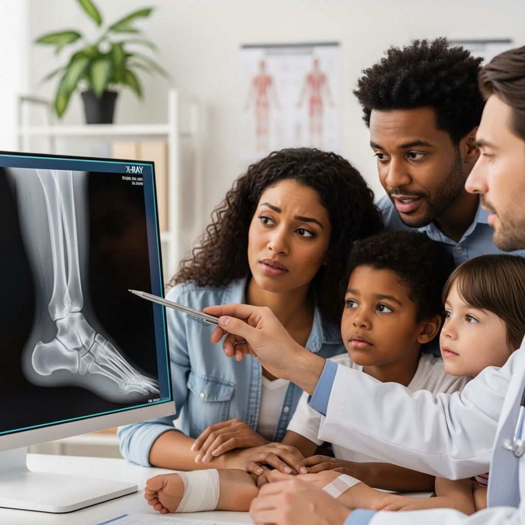
When a child twists, falls on, or suddenly refuses to put weight on an ankle, an X-ray is often the quickest way for doctors to see whether a bone is broken or if there is another cause for the pain. An ankle X-ray is a fast, painless imaging test that creates pictures of the bones in the lower leg and ankle using a very small amount of radiation. Understanding what will happen and why it is being done can help parents keep children calm, informed, and reassured.
Why Doctors Recommend Ankle X-Rays
Doctors do not order ankle X-rays for every bump or twist. Instead, they use the child’s symptoms, the way the injury happened, and a physical exam to decide whether an image is needed. The main goal is to determine whether there is a fracture, a severe sprain, or another serious problem that might require a cast, brace, or surgery.
- To check for fractures, such as small cracks or complete breaks in the bones around the ankle.
- To evaluate severe sprains, in which ligaments may be stretched or torn and might pull off a small piece of bone.
- To rule out serious injury when a child has significant pain, swelling, or trouble walking after a fall or sports injury.
- To monitor healing after treatment, such as checking alignment after a cast or surgery.
Sometimes, clinical decision rules help doctors decide whether an X-ray is really necessary. These guidelines focus on where the child feels pain, whether certain spots on the ankle are tender to touch, and if the child can stand or walk a few steps. If the risk of a fracture seems low, the doctor may choose rest and observation instead of an X-ray.
Basic Ankle Anatomy for Parents
Knowing a little about the ankle’s structure makes it easier to understand the X-ray report and your child’s diagnosis. The ankle is a complex joint where the leg bones meet the foot, allowing motion such as pointing the toes up and down or turning the foot inward and outward.
- Tibia: The larger bone of the lower leg, often called the shin bone, which forms the inner part of the ankle.
- Fibula: The thinner bone on the outer side of the lower leg that forms the outer ankle bump.
- Talus: The bone that sits under the tibia and fibula, acting as the main link between the leg and the foot.
- Growth plates: Soft areas of growing tissue near the ends of a child’s bones that appear differently from fully formed bone on an X-ray.
- Ligaments: Strong bands of tissue that connect bones and stabilize the ankle joint, often injured in sprains.
Your child’s X-ray focuses on this area to show whether the bones are intact, aligned properly, and growing as expected.
Different Types of Ankle X-Ray Views
An ankle X-ray is usually a set of images taken from different angles, not just a single picture. Taking more than one view helps ensure that small fractures or changes in alignment are not hidden by overlapping bones.
| View name | How the ankle is positioned | What this view helps show |
|---|---|---|
| Front (AP) view | Foot facing forward, ankle straight | Overall look at the tibia, fibula, and talus from the front |
| Angle (oblique) view | Foot turned slightly inward | Clearer view of the ankle joint space where the bones meet |
| Side (lateral) view | Foot turned so the outside edge rests on the table | Side view of the ankle, heel bone, and back of the foot |
In some cases, the healthcare provider may request additional views or images of the foot or lower leg if symptoms suggest that the injury extends beyond the ankle area.
Preparing a Child for an Ankle X-Ray
Most children do not need special medical preparation for an ankle X-ray, but emotional preparation makes a big difference. A calm explanation in simple words helps reduce fear, especially if a child has never had an X-ray before.
- Explain that the test uses an invisible “camera” that can see bones, and that it does not hurt.
- Reassure your child that nothing will touch or poke the ankle during the picture, although the technologist may gently move the leg.
- Let your child know that staying still for a few seconds at a time helps the images come out clearly and avoids needing extra pictures.
Comfort items, such as a favorite toy, blanket, or stuffed animal, are often allowed in the room as long as they do not interfere with the imaging. Parents are usually invited to remain nearby for reassurance, though they may be asked to wear a protective apron.
What Happens During the Appointment
The actual X-ray process is usually quick, often just a few minutes once your child is in the imaging room. The longer part of the visit is often registration and waiting for the images to be ready for the doctor to review.
- Check-in and brief history: A staff member may ask about how the injury happened, where it hurts, and whether your child has had X-rays before.
- Positioning on the table: Your child may be asked to lie down, sit, or occasionally stand on a step, depending on the views required and how much the ankle hurts.
- Protective shields: A lead apron or shield might be used to cover parts of the body that do not need imaging.
- Taking the images: The technologist moves the machine close to the ankle, steps behind a barrier, and asks your child to hold still while the images are taken.
- Repositioning: The ankle is gently moved for each view, with pillows or foam pads used to support a painful limb whenever possible.
There is no sensation from the X-rays themselves. The only discomfort your child may feel usually comes from moving a sore ankle into different positions, which the technologist tries to minimize.
Radiation and Safety Considerations
Parents often worry about radiation exposure from imaging tests. An ankle X-ray uses a very low dose, far below the levels linked with health problems, especially when used occasionally and for a specific medical reason. The benefits of detecting or ruling out a serious injury almost always outweigh the minimal risk from a single test.
- Only the area of interest, in this case the ankle and nearby bones, is exposed to the X-ray beam.
- Protective shields and focused beams help reduce exposure to the rest of the body.
- Technologists are trained to use the smallest dose that will still produce clear, usable images.
Children are more sensitive to radiation than adults over a lifetime, which is why medical teams are careful to limit imaging to what is truly needed. If you have questions about the dose or other imaging options, your child’s doctor or the radiology team can explain the risks and benefits in more detail.
What Ankle X-Rays Can and Cannot Show
An X-ray is excellent for showing bones, but it has limits. Bone appears bright on the image, while soft tissues such as muscles, tendons, and ligaments are less visible. This is why many injuries are diagnosed based on both the X-ray and the physical exam.
- Conditions commonly seen on ankle X-rays:
- Small or large fractures of the tibia, fibula, or bones of the foot near the ankle.
- Growth plate injuries that may affect how a bone grows if not handled correctly.
- Bone alignment problems, such as bones that are out of place after a fracture.
- Conditions that may not show clearly:
- Minor ligament sprains and tendon injuries.
- Very small cracks or stress fractures early on, which sometimes appear only after healing begins.
If your child continues to have significant pain despite a normal X-ray, the doctor may repeat imaging later or consider other tests such as an MRI or ultrasound to look more closely at soft tissues.
Understanding the Results and Next Steps
After the images are taken, a radiologist, who is a doctor specialized in interpreting medical images, reviews them and sends a report to the healthcare provider who ordered the test. In emergency situations, preliminary findings are often shared quickly so treatment can begin without long delays.
| Possible X-ray finding | What it means | Typical next steps |
|---|---|---|
| Normal bones, no fracture | No visible break or misalignment | Rest, ice, support bandage, and follow-up if pain persists |
| Simple fracture in good position | Clean break with bone ends lined up | Cast, boot, or splint; limited weight-bearing until healed |
| Displaced or complex fracture | Bone pieces out of position or multiple fragments | Possible realignment, cast, or surgery; closer follow-up |
| Growth plate involvement | Injury includes the area where the bone grows | Careful monitoring; sometimes specialist referral |
Ask the provider to explain the findings in simple language and to show you the images if possible. Seeing the picture often makes it easier to grasp why a cast, brace, or specific activity restrictions are needed.
Helping Your Child Cope Before and During the X-Ray
Children react to medical tests in different ways, depending on age, temperament, and past experiences. Some may be curious, while others are fearful or resistant. Parental support can make the experience smoother and less stressful.
- Use age-appropriate language, such as “bone pictures” or “special camera,” instead of technical terms that may sound frightening.
- Practice staying still at home by playing a “statue game” to show how holding still is part of the test.
- Stay close to your child’s side, holding a hand or talking softly if allowed in the room.
- Plan a small reward afterward, such as a story, a favorite activity, or a special snack, to give your child something positive to anticipate.
For very young or very anxious children, the team may take a little extra time to explain the equipment, let them touch safe parts of the machine, or show how the table moves to reduce fear of the unknown.
After the X-Ray: Care and Monitoring
Once the X-ray is finished, your child can usually return to normal activities right away, unless the healthcare provider recommends otherwise because of the injury itself. There are no lingering effects from the imaging, and no special precautions are needed beyond the treatment plan for the ankle.
- Follow instructions about weight-bearing, whether your child can walk on the ankle or should use crutches.
- Use ice, elevation, and pain relief medicines as directed to control swelling and discomfort.
- Watch for warning signs such as increasing pain, numbness, or color changes in the toes, which warrant a prompt call to the doctor.
Follow-up visits or repeat X-rays may be scheduled to check how well a fracture is healing or to ensure that the bones stay in good alignment while your child grows.
Common Misconceptions About Ankle X-Rays
Misunderstandings about imaging tests can add unnecessary worry. Clarifying what X-rays can and cannot do helps parents make informed decisions and ask the right questions at the clinic.
- An X-ray is not a treatment; it is a tool used to diagnose and guide treatment decisions.
- A normal X-ray does not mean your child is “faking it” or that the pain is not real; soft tissue injuries can hurt just as much as fractures.
- Needing more images does not always mean something is wrong; sometimes views are repeated simply to improve clarity.
- One ankle X-ray does not significantly increase long-term cancer risk when proper safety measures are used.
When to Seek Urgent Care for an Ankle Injury
Many minor ankle injuries can be handled at home with rest and careful observation. However, certain signs mean a child should be evaluated quickly, and an X-ray may be part of that assessment.
- Visible deformity of the ankle or foot, such as a crooked or twisted appearance.
- Inability to stand or take even a few steps without severe pain.
- Rapid swelling or bruising, especially if combined with intense tenderness over the bone.
- Open wounds near the ankle, or bone that appears to be pushing against or through the skin.
- Changes in skin color, temperature, or sensation in the foot or toes.
If any of these are present, call your child’s healthcare provider or visit an urgent care or emergency department. Quick evaluation helps prevent complications and ensures that serious injuries are not missed.
FAQs About Ankle X-Rays in Children
Does an ankle X-ray hurt?
The X-ray itself is painless because it is simply a type of light that passes through the body. Any discomfort your child feels usually comes from moving or positioning a sore ankle, which the technologist will try to do as gently as possible.
How long does the whole process take?
The actual imaging usually takes only a few minutes, but the total time in the department may be longer because of registration, waiting, and reviewing results. Plan for at least 30–60 minutes at the facility, especially in a busy emergency setting.
Can I stay with my child during the X-ray?
In most cases, a parent or caregiver is allowed to stay in the room as long as safety rules are followed. You may be asked to wear a protective apron and stand in a specific spot while the images are taken.
Will my child need sedation?
For a standard ankle X-ray, sedation is rarely needed. Most children, even toddlers, can complete the test with gentle support and clear instructions. Sedation is more common for longer imaging tests, such as certain types of MRI, where staying completely still for a long period is necessary.
How will we get the results?
In an emergency or urgent care setting, the doctor who ordered the X-ray usually discusses the findings with you before you leave. In other offices, results may be shared during a follow-up visit, by phone, or through an online patient portal, depending on local practices.
Are there alternatives to an X-ray?
For bone injuries, X-rays remain the main first-line test because they are quick, widely available, and effective. Other studies, such as ultrasound, CT scans, or MRI, may be used in special situations but are usually reserved for more complex or unclear cases.
Key Takeaways for Parents
- An ankle X-ray is a safe, quick way to check for broken bones or serious injury when a child has significant ankle pain, swelling, or trouble walking.
- The test does not hurt, uses a very small amount of radiation, and is performed by trained staff who focus on minimizing exposure.
- Staying calm, explaining the process in simple terms, and offering comfort and reassurance can greatly improve your child’s experience.
- Understanding the results and following treatment instructions carefully help protect your child’s growing bones and support a smooth recovery.
With the right information and support, parents can approach an ankle X-ray with confidence, knowing it is an important tool to keep a child’s active feet and ankles healthy and strong.