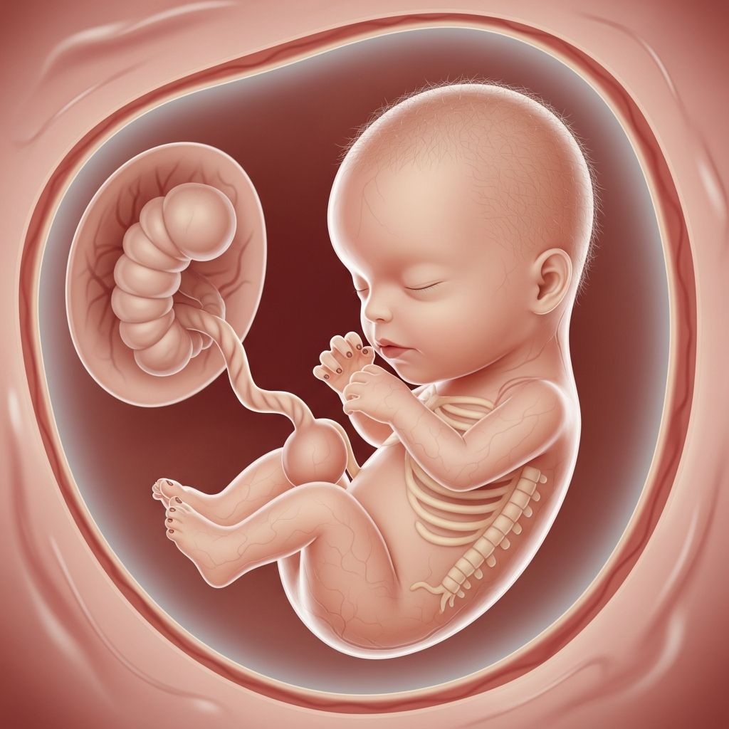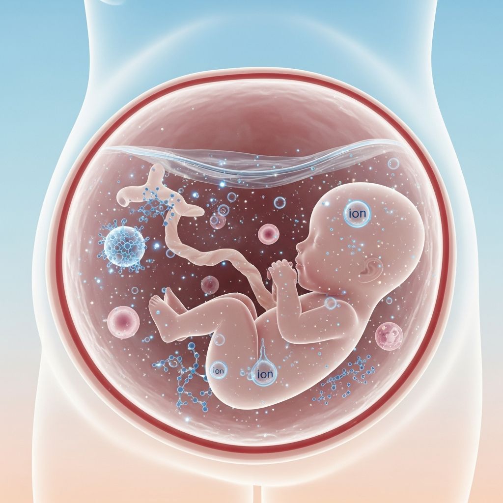
Amniotic fluid serves as a vital lifeline for the developing fetus, providing a protective environment within the amniotic sac. This dynamic fluid, primarily composed of water and electrolytes, evolves in content and volume throughout gestation to support growth, movement, and organ maturation.
The Formation and Sources of Amniotic Fluid
Amniotic fluid begins accumulating shortly after the gestational sac forms, initially derived from maternal plasma that diffuses across fetal membranes via osmotic and hydrostatic pressures. In the early embryonic phase, up to around 10-12 weeks, the fluid resembles a protein-poor dialysate of maternal and fetal plasma, entering through the permeable fetal skin and amniotic epithelium.
As pregnancy advances into the second trimester, fetal kidneys activate around week 16, shifting the primary source to fetal urine, which becomes hypotonic and contributes progressively more volume. By late gestation, fetal urine accounts for 500-700 ml daily, supplemented by lung liquid secretions (up to one-third of total fluid), gastrointestinal outputs, and minor umbilical cord or placental contributions.
Fluid dynamics involve a balance of production and removal: fetuses swallow 200-450 ml/day and intramembranous absorption occurs across the amnion into fetal vessels, maintaining equilibrium. This bidirectional exchange ensures the fluid remains hypotonic (250-260 mOsm/L near term) compared to fetal plasma.
Biochemical Makeup of Amniotic Fluid
At its core, amniotic fluid is 98% water with electrolytes like sodium, chloride, and potassium mirroring maternal serum levels. The remaining 2% includes signaling molecules, peptides, carbohydrates, lipids, hormones, urea, creatinine, uric acid, and extracellular matrix components such as collagens, glycosaminoglycans (hyaluronic acid, chondroitin sulfate), proteins, phospholipids, and fetal-derived biochemicals.
Early in pregnancy, the fluid is clear and less viscous due to low protein content. By 12-14 weeks, it incorporates nutrients essential for fetal growth, infection-fighting antibodies, and hormones aiding organ development. The pH ranges from 7.0 to 7.5, contrasting with the acidic vaginal environment (pH 3.8-4.5), which helps diagnose membrane rupture via pH testing.
| Component | Early Pregnancy | Late Pregnancy | Function |
|---|---|---|---|
| Water & Electrolytes | Primary (98%) | Primary (98%) | Hydration, osmotic balance |
| Fetal Urine | Minimal | 500-700 ml/day | Main volume source |
| Lung Secretions | Minor | ~1/3 of volume | Lung maturation |
| Proteins/Lipids/Carbs | Trace | Increased | Fetal nutrition, growth |
| pH | Neutral | 7.0-7.5 | Stable environment |
Volume Dynamics Across Gestational Stages
Amniotic fluid volume (AFV) correlates closely with fetal size in the first half of pregnancy. At 10 weeks, it measures about 25-60 ml, rising to 175 ml by 16 weeks and 400 ml by 20 weeks. Peak volume of 800-1200 ml occurs at 34-38 weeks, then declines to 600-800 ml at term (40 weeks) and further post-term.
- Early (8-20 weeks): Dialysis via fetal skin dominates; steady rise tied to fetal weight.
- Mid (20-34 weeks): Urine production surges; AFV expands rapidly with fetal breathing/swallowing influencing balance.
- Late (34+ weeks): Plateau then decline; reduced urine output post-40 weeks, increased absorption.
Clinicians assess AFV using Amniotic Fluid Index (AFI) via ultrasound, with normal ranges varying by week (e.g., mean AFI peaks mid-third trimester). Factors like fetal swallowing (keratinized skin post-25 weeks) and lung fluid exchange fine-tune levels.
Critical Functions in Fetal Protection and Development
Beyond cushioning against uterine compression, amniotic fluid maintains constant temperature and pressure, allowing free fetal movement for musculoskeletal development. It facilitates lung maturation through fetal ‘breathing’ motions, practicing inhalation/exhalation in a liquid medium.
The fluid supports gastrointestinal practice via swallowing, aids ear and urinary tract development, and provides biochemical exchange with the mother, including antibodies for immunity. Nutrients and hormones promote organ growth, while its clarity and composition prevent infections and promote skin protection via vernix caseosa flakes.
Recognizing Normal vs. Abnormal Characteristics
Healthy amniotic fluid appears clear or light yellow, odorless or mildly sweet, with minimal vernix particles. Color changes signal issues: green/brown indicates meconium passage (fetal distress); yellow tinges may suggest bilirubin; cloudy fluid could mean infection.
Volume imbalances pose risks:
- Oligohydramnios (low AFV): <5th percentile AFI; linked to renal agenesis, membrane leaks, or post-term pregnancy. Risks: pulmonary hypoplasia, limb deformities, cord compression.
- Polyhydramnios (high AFV): >95th percentile; caused by fetal swallowing issues (GI atresia), anencephaly, maternal diabetes. Risks: preterm labor, abruption, malpresentation.
Clinical Monitoring and Diagnostic Insights
Ultrasound measures AFV serially, especially in high-risk cases. Amniocentesis samples fluid for genetic testing, lung maturity (L/S ratio), or infection markers. Vaginal pH >4.5 with clear discharge suggests rupture; ferning test or nitrazine confirms.
Understanding fluid dynamics informs interventions: amnioinfusion for oligohydramnios, early delivery for severe polyhydramnios. Recent studies emphasize AFV’s predictive value for perinatal outcomes.
Frequently Asked Questions (FAQs)
What does amniotic fluid look and smell like normally?
Clear to light yellow, odorless or faintly sweet, with possible vernix specks. Abnormal colors like green signal potential distress.
How much amniotic fluid is normal at term?
About 600-800 ml at 40 weeks, peaking earlier at 800-1200 ml.
Can amniotic fluid levels change suddenly?
Yes, leaks from membrane rupture or imbalances from fetal/maternal conditions can alter levels rapidly, requiring monitoring.
Does low amniotic fluid always harm the baby?
Not always; mild cases may resolve, but severe/prolonged oligohydramnios risks lung and limb issues.
What causes excess amniotic fluid?
Often fetal anomalies impairing swallowing or maternal conditions like diabetes.
References
- Embryology, Amniotic Fluid — StatPearls, NCBI Bookshelf. 2023-11-03. https://www.ncbi.nlm.nih.gov/books/NBK541089/
- Amniotic fluid — Wikipedia. 2024-01-12. https://en.wikipedia.org/wiki/Amniotic_fluid
- Amniotic Fluid: Physiology and Assessment — GLOWM. 2023. https://www.glowm.com/section-view/heading/Amniotic%20Fluid:%20Physiology%20and%20Assessment/item/208
- Amniotic fluid — MedlinePlus Medical Encyclopedia. 2024-05-01. https://medlineplus.gov/ency/article/002220.htm
- Amniotic fluid: Functions and disorders — Medical News Today. 2023-07-15. https://www.medicalnewstoday.com/articles/307082
- Amniotic fluid characteristics and its application in stem cell therapy — PMC. 2022-10-01. https://pmc.ncbi.nlm.nih.gov/articles/PMC9596929/