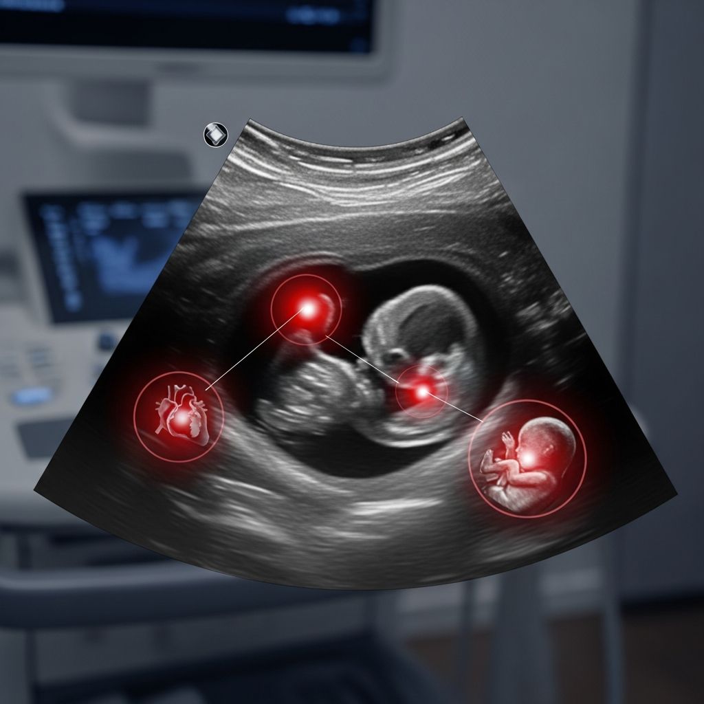
Fetal ultrasounds provide vital insights into your baby’s development, but certain findings can raise alarms requiring prompt attention. These scans use sound waves to create images, helping detect potential problems early, though not all irregularities indicate severe issues.
Understanding the Role of Ultrasound in Prenatal Monitoring
Prenatal ultrasounds are standard in most pregnancies, typically performed at 8-14 weeks for dating and viability, 18-22 weeks for anatomy, and later for growth checks. They assess heartbeat, position, organs, and placenta, offering reassurance or flagging concerns like delayed growth or malformations.
While generally safe with low risks, ultrasounds involve energy exposure that warrants prudent use, especially avoiding non-medical ‘keepsake’ scans. Organizations like the Mayo Clinic emphasize medical necessity to minimize any theoretical bioeffects.
Common Abnormalities Detected on Scans
Several ultrasound features can signal trouble. Here’s a breakdown:
- Absent or Irregular Heartbeat: In early scans, no heartbeat by 7-10 weeks may indicate miscarriage risk, though follow-up confirms viability.
- Excessive Nuchal Fluid: Thickened nuchal translucency (>3mm) at 11-14 weeks raises chromosomal abnormality risks, but 19/20 high-risk cases resolve normally.
- Short or Absent Nasal Bone: Linked to Down syndrome, often combined with other markers.
- Echogenic Intracardiac Focus: Bright spots in the heart, a soft marker for trisomies, usually benign alone.
These markers prompt further tests like noninvasive prenatal testing (NIPT) or amniocentesis for clarity.
Growth-Related Concerns in Mid-to-Late Pregnancy
By the anatomy scan, growth discrepancies emerge:
| Issue | Ultrasound Sign | Possible Causes |
|---|---|---|
| Intrauterine Growth Restriction (IUGR) | Small measurements (<10th percentile) | Placental insufficiency, maternal hypertension |
| Macrosomia | Large estimates (>90th percentile) | Gestational diabetes, obesity |
| Oligohydramnios/Polyhydramnios | Low/High Amniotic Fluid Index | Kidney issues, swallowing problems/swallowing excess |
Amniotic fluid imbalances are critical; low levels may signal fetal kidney distress, while excess could indicate gut blockages. Serial scans monitor these.
Structural Defects and Organ Anomalies
Serious red flags include organ malformations:
- Neural Tube Defects: Anencephaly (missing skull/brain) or spina bifida (spinal opening) appear as absent cranial vault or ‘lemon’ skull sign.
- Heart Defects: Holes, valve issues, or single ventricle show abnormal four-chamber views.
- Limb Abnormalities: Missing or short bones, clubfoot.
- Cleft Lip/Palate: Facial clefts visible in 3D/2D profiles.
These affect 2-3% of pregnancies; early detection allows planning, sometimes fetal surgery.
Placental and Umbilical Cord Warning Signs
Placenta problems pose risks:
- Placenta Previa: Covers cervix, risking bleeding; monitored closely.
- Placental Abruption: Separation shows retroplacental hematoma.
- Vasa Previa: Cord vessels over cervix, dangerous at delivery.
Cord issues like single artery or knots may indicate chromosomal problems or growth restriction. Doppler checks blood flow; absent end-diastolic flow signals severe IUGR.
Position and Presentation Issues Late in Pregnancy
After 32 weeks:
- Breech Position: Bottom-first; may need version or C-section.
- Transverse Lie: Sideways, complicating vaginal birth.
- Umbilical Cord Prolapse: Rare but emergency if membranes rupture.
These often resolve, but persistent cases require intervention.
Interpreting ‘Soft Markers’ vs. Hard Indicators
Distinguish subtle from overt issues:
| Type | Examples | Risk Level | Action |
|---|---|---|---|
| Soft Markers | Choroid plexus cyst, echogenic bowel | Low alone; reassess | Genetic counseling if multiple |
| Hard Indicators | Holoprosencephaly, omphalocele | High; often lethal | Amnio, MRI, specialist referral |
Soft markers like cysts vanish by 26 weeks without issue.
The Debate on Ultrasound Safety and Potential Risks
Ultrasounds are deemed safe by WHO reviews, showing no links to birth defects, preterm birth, or neurodevelopmental delays in humans. Animal studies note thermal/mechanical effects at high exposures, but human routine scans (under 15 min) don’t elevate temperature sufficiently.
Subtle associations exist with non-right-handedness in boys or speech delays, but evidence is weak and confounded. ACOG and AIUM advocate ALARA principle: minimal exposure for medical benefit, avoiding entertainment scans. PubMed reviews affirm low risks but note bioeffects potential.
What to Do If Your Scan Shows Concerns
- Stay Calm: Many findings are false positives; discuss probabilities with your doctor.
- Seek Details: Ask for images, measurements, and risk stats.
- Pursue Follow-Ups: Repeat scans, specialist consults (maternal-fetal medicine).
- Consider Diagnostics: NIPT, CVS, amnio for genetics; MRI for brain/spine.
- Plan Delivery: NICU prep, C-section if needed.
Multidisciplinary teams guide decisions, including termination options if severe.
Advances in Ultrasound Technology Enhancing Detection
3D/4D imaging clarifies anomalies, Doppler assesses flows precisely. Fetal echocardiography detects 70% heart defects missed in standard scans. AI aids measurements, reducing errors.
Frequently Asked Questions
Can a bad ultrasound mean my baby isn’t healthy?
Not always; many abnormalities resolve, and false positives occur. Follow-up clarifies.
Are ultrasounds completely safe?
Low risk per WHO and Mayo Clinic; use medically, follow ALARA.
What if fluid levels are abnormal?
Monitor closely; low fluid may need amnioinfusion, high fluid swallowing checks.
How accurate are growth predictions?
±15%; late scans better. Serial checks essential.
Should I worry about soft markers?
Usually no if isolated and baby otherwise normal; genetic tests if concerned.
Empowering Expecting Parents with Knowledge
Ultrasound anomalies can be daunting, but informed parents partner effectively with providers. Early detection improves outcomes, from simple monitoring to interventions. Trust verified processes, seek second opinions, and focus on holistic care.
References
- Ultrasound Scans- Cause for Concern — Dr. Sarah Buckley. Accessed 2026. https://sarahbuckley.com/ultrasound-scans-cause-for-concern/
- Effects of Ultrasound on Biological Tissues and Cells — Global Library of Women’s Medicine (GLOWM). Accessed 2026. https://www.glowm.com/article/heading/vol-5–surveillance-of-fetal-wellbeing–effects-of-ultrasound-on-biological-tissues-and-cells/id/411403
- Are Too Many Ultrasounds Harmful? Experts Weigh In — CCRM Fertility. Accessed 2026. https://www.ccrmivf.com/news/fertility-crisis-flint-2/
- Benefits and risks of ultrasound in pregnancy — PubMed (NCBI). 2013-10-26. https://pubmed.ncbi.nlm.nih.gov/24176149/
- Why to avoid ‘keepsake’ 3-D and 4-D ultrasounds — UT Southwestern Medical Center. Accessed 2026. https://utswmed.org/medblog/3d-4d-ultrasound/
- Fetal ultrasound — Mayo Clinic (.org). Accessed 2026. https://www.mayoclinic.org/healthy-lifestyle/pregnancy-week-by-week/in-depth/fetal-ultrasound/art-20546827
- Safety of ultrasonography in pregnancy: WHO systematic review — Ultrasound in Obstetrics & Gynecology (Wiley). Accessed 2026. https://obgyn.onlinelibrary.wiley.com/doi/full/10.1002/uog.6328