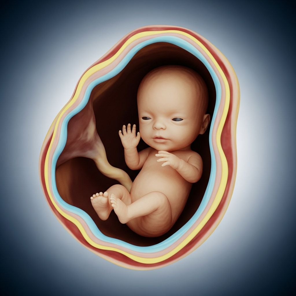
Three-dimensional (3D) ultrasounds represent an advanced imaging technique in prenatal care, offering detailed views of the fetus beyond traditional two-dimensional (2D) scans. While they captivate many expectant parents with lifelike images, their role is primarily medical rather than recreational. This article delves into the technology, its applications, benefits, risks, and guidelines from leading health authorities to help you make informed decisions.
What Are 3D Ultrasounds and How Do They Work?
3D ultrasounds use high-frequency sound waves to create volumetric images of the fetus, reconstructing flat 2D slices into a three-dimensional model. This process involves specialized transducers that capture multiple angles simultaneously, allowing for surface rendering of facial features, limbs, and organs. Unlike 2D ultrasounds, which provide cross-sectional views, 3D imaging enables rotation and manipulation of the image post-scan for better visualization.
The technology builds on standard ultrasound principles but requires more processing power and skilled operators. Four-dimensional (4D) ultrasounds extend this by adding real-time motion, showing the baby moving. These scans typically occur between 24-32 weeks of gestation when amniotic fluid is ample and fetal features are well-formed.
- Key components: Transducer probe, computer software for 3D reconstruction, and gel for sound wave transmission.
- Scan duration: 20-45 minutes, longer than routine 2D scans due to image optimization.
- Settings: Performed in low-light rooms for optimal probe handling and maternal comfort.
Medical Applications: When 3D Ultrasounds Shine
In clinical practice, 3D ultrasounds serve as a problem-solving tool when 2D scans yield inconclusive results. They excel in evaluating complex anatomical structures, particularly in high-risk pregnancies. For instance, they enhance detection of facial clefts, where 2D images may miss subtle palate defects by providing multi-planar views.
Other critical uses include:
- Assessing skeletal dysplasias through precise bone measurements.
- Evaluating neural tube defects like spina bifida via surface rendering.
- Monitoring twins or multiples for growth discrepancies and shared placenta issues.
- Investigating cardiac anomalies with volume datasets for offline analysis.
Studies show 3D ultrasound reduces operator dependency, making it valuable for telemedicine in remote areas. In high-risk cases, such as suspected preterm delivery, it has outperformed 2D in predicting outcomes.
Comparing 2D, 3D, and 4D Ultrasounds
To clarify differences, consider this comparison table:
| Feature | 2D Ultrasound | 3D Ultrasound | 4D Ultrasound |
|---|---|---|---|
| Image Type | 2D cross-sections | 3D static volumes | 3D in real-time motion |
| Primary Use | Routine screening | Diagnostic detail | Dynamic assessment |
| Time per Scan | 10-20 min | 20-45 min | 30+ min |
| Cost | Standard (covered) | Higher (often elective) | Highest |
| Best For | Heartbeat, position | Anomaly detection | Fetal behavior |
2D remains the gold standard for most prenatal visits due to its speed, cost-effectiveness, and proven safety profile.
Potential Benefits for Expectant Parents
Beyond diagnostics, 3D images foster emotional connections. Seeing a detailed face can strengthen parental bonding, potentially reducing anxiety in uncertain pregnancies. Some research suggests this visualization aids psychological preparation, though randomized trials are limited.
In medical contexts, clearer images empower informed discussions about management plans, such as surgical interventions for cleft repairs post-birth. For families with prior losses, these scans provide reassurance when anomalies are ruled out.
Safety Concerns and Potential Risks
Ultrasound is noninvasive, but non-medical “keepsake” 3D/4D scans raise safety flags. The FDA and ACOG caution against elective use, citing unknown long-term effects of prolonged exposure. Ultrasound generates heat via tissue absorption, adhering to the ALARA principle (As Low As Reasonably Achievable) to minimize energy.
Reported risks include:
- Thermal effects: Elevated fetal temperature linked to potential birth defects in animal studies.
- False results: Artifacts mimicking pathologies, like apparent limb defects from shadows.
- Developmental concerns: Associations with low birth weight, speech delays, or non-right-handedness in some epidemiological data, though causality is unproven.
- Oligohydramnios challenges: Poor image quality in low fluid, leading to misinterpretation.
The AIUM notes pressures for bonding scans but urges prudent use, as 3D adds time and exposure without routine medical gain.
Expert Guidelines from Leading Organizations
Authoritative bodies provide clear stances:
- FDA: Discourages non-diagnostic fetal imaging; ultrasound is safe when medically indicated.
- ACOG: Endorses AIUM’s prudent approach, limiting to necessary diagnostics.
- AIUM: No confirmed harm from diagnostic use, but cautions on entertainment scans.
- WHO: Deems diagnostic ultrasound safe based on evidence, but not for souvenirs.
These align: Reserve 3D for cases where it alters care, performed by certified sonographers.
Cost, Availability, and Insurance Coverage
3D ultrasounds cost $100-$400 out-of-pocket, as insurance covers only medically necessary scans. Boutique studios offer packages, but lack medical oversight. Availability varies; hospitals provide them diagnostically, while private centers focus on electives. Always verify technician credentials (RDMS certification).
Alternatives to 3D Ultrasounds
For bonding without risks:
- Standard 2D anatomy scans at 18-22 weeks.
- Fetal Doppler for heartbeat listening at home.
- Maternity photos or apps simulating fetal growth.
- Post-birth professional photography.
Advanced MRI serves for complex diagnostics without sound waves.
Frequently Asked Questions (FAQs)
Is a 3D ultrasound safe for my baby?
Diagnostic 3D ultrasounds are considered safe under medical supervision, following ALARA. Avoid elective scans to minimize exposure.
Can 3D ultrasounds detect all birth defects?
No; they complement 2D but miss issues like chromosomal abnormalities requiring genetic tests.
When is the best time for a 3D scan?
24-32 weeks, when facial features are prominent and fluid supports imaging.
Are 3D ultrasounds covered by insurance?
Only if medically necessary; elective ones are not.
Do 3D scans improve pregnancy outcomes?
In targeted cases yes, by refining diagnoses; routinely, 2D suffices.
Making the Right Choice for Your Pregnancy
Discuss with your obstetrician whether 3D adds value. Prioritize evidence-based care over novelty. Routine prenatal visits with 2D ultrasounds, genetic screening, and lifestyle factors ensure the best outcomes.
References
- Are 3D ultrasounds recommended? — Premier Care For Women. Accessed 2026. https://premiercare4womenaz.com/are-3d-ultrasounds-recommended/
- The Risks And Benefits Of 3D/4D Ultrasound Scans For Prenatal Diagnosis — 3D4D Studio. Accessed 2026. https://3d4dstudio.com/the-risks-and-benefits-of-3d-4d-ultrasound-scans-for-prenatal-diagnosis/
- Against – 3D ultrasound in first and second trimester pregnancy — PMC (NCBI). 2016-09-13. https://pmc.ncbi.nlm.nih.gov/articles/PMC5024843/
- Benefits and Risk of Fetal 3D Ultrasound — Allied Health CE Connection. Accessed 2026. https://alliedhealth.ceconnection.com/files/PicturePerfect-1346101146862.pdf
- Why to avoid ‘keepsake’ 3-D and 4-D ultrasounds — UT Southwestern Medical Center. Accessed 2026. https://utswmed.org/medblog/3d-4d-ultrasound/
- Prudent Use and Safety of Diagnostic Ultrasound in Pregnancy — AIUM. 2020-05-19. https://www.aium.org/resources/official-statements/view/prudent-use-and-safety-of-diagnostic-ultrasound-in-pregnancy
- Use of 3-D, 4-D or 5-D Ultrasound in Maternity Care — Healthy Blue NC Provider. Accessed 2026. https://provider.healthybluenc.com/medpolicies/healthybluenc/active/mp_pw_a053265.html
- Ultrasound Imaging — FDA. Accessed 2026. https://www.fda.gov/radiation-emitting-products/medical-imaging/ultrasound-imaging